Infectious Disease Update: Clinical Pearls

Ted Rosen, MD

At MauiDerm Fall NP+PA 2014, Dr Rosen provides us with an update on infectious diseases and his clinical pearls…

  1. STDs are globally epidemic. In the US, 50% of those 15-25 have or have had an STD
  2. STDs are so common due to the “multiplier” effect of sex partners of sex partners
  3. Syphilis is on the rise last three years in USA, now at 5.3/100,000 population
  4. Multi-drug resistant GC (including resistance to drugs of choice, cephalosporins) is here
  5. Topical three months regimen of 4% zinc sulfate soaks for active genital herpes benficial
  6. External genital warts: NEW treatment ideas
    • 5% KOH daily x 3 months
    • Single application of ingenol mebutate (either strength)
    • Thermotherapy with 440C for 30 min x 3 days, each of two consecutive weeks
  7. Leishmaniasis being brought back to USA from Iraq and Afghanistan
    • Rx: Pentavalent antimonial IV, IM from CDC; Quite toxic.
    • Thermotherapy (ThermoMed device): 1300F; Painful and blisters
    • New drug: Miltefosine 100-150mg/day PO (Impavido™) available 2015
      • Lecithin derivative
      • Safe and effective for Old and New World Leishmaniasis
  8. Treatments for MRSA have expanded with three new drugs
    • All new drugs have LONG T1/2 which allows less frequent dosing
    • Oritivancin: Single 1200mg dose
    • Dalbavancin: 1000mg, then 500mg a week later
    • Tedizolid: 200mg QD x 5-7 days
  9. Chikungunya virus
    • Used to be Africa/Asia, Now worldwide
    • Most cases imported from trip (especially from Caribbean)
    • Now, virus-carrying mosquitoes in Caribbean and USA
    • Fever, arthralgia, myalgia, headache, petechial to purpuric rash
    • Like Dengue but not as bad
    • No vaccination, No specific Rx. Supportive therapy, as needed.

Cutaneous Oncology: Clinical Pearls

George Martin, MD

How are you managing your cutaneous oncology patients? Dr Martin provides us with his clinical pearls…

  • Remember that AK patients are not just Medicare patients, we see patients 30-40 years old as well. Also remind older patients that actinic damage is chronic disease and no one therapy will result in a long lasting or permanent cure.
  • When treating AKs, expect side effects based on the MOA of the active ingredient.
  • Treating AKs on the chest? Therapeutic considerations include:  Topical 5-FU cream 0.5/1.5%; Ingenol Mebutate gel 0.5%; and Diclofenac gel 3%., Expect a significant skin reaction to ingenol mebutate 0.05% (not .5%) within 24 hrs that will include blistering and discomfort requiring analgesia when large areas are covered.. 5-FU can be “titrated” and can be used for 7-10  days, discontinued for a month and re-started in a month and used for 2-3 weeks with less of a reaction. DO NOT USE IMIQUIMOD on the chest as it is not FDA approved for the chest and  an result in permanent depigmentation.
  • Managing reactions is key! Utilize analgesia as needed; sunscreen to reduce potential for dyschromia; moisturizers; topical antibiotics as needed; and weekly follow-up.
  • Imiquimod can trigger flu-like symptoms including myalgias and fever before a significant skin reaction such as erosions and ulceration may occur.
  • Ingenol Mebutate has two mechanisms of action: an immediate cytotoxic effect initiated via the PKC pathway which causes swelling  and edema even blistering and weeping and a second MOA involving IL-8 production by rapidly proliferating ketatinocytes and endothelial cells which recruits neutrophils and in vigorous responses can result in sterile pustules.
  •  Counsel patients on anticipated responses and set expectations.
  • Have patients fill prescriptions between Monday to Thursday—less likely to be switched than Fridays or weekends.
  • Have patients start treatments on Sundays, for ingenol mebutate during Mon-Thurs as the immediate reaction to ingenol mebutate may trigger a phone call within 24 hours; this way, reactions occur mid week rather than on weekends.
  • Make sure that surgical sites are well healed and that cryotherapy sites have had a week or two after healing before initiating therapy.
  • Use every adjunct possible except steroids as the immune system plays a role in each modality used.

Social Media

Jeff Benabio, MD

At MauiDerm 2014, Dr Benabio provided an overview and update on Social Media and its potential use in clinical practice…

Did you know that Yelp is a social media website? It is and for doctors, this is something that you should know. Why does the New York Times pose questions at the bottom of their posts? They do this in order to engage their customers and it turns out then when people participate in your brand, the lifetime value of that customer goes up. This is very much what is happening with social media.

The What, Why and How of Social Media

Remember that social media is a lot more than just Facebook! Social media is digital technologies that allow us to connect and share things/ideas with each other. This is a core human capability, i.e., connecting with people. The internet, in its entirety, is essentially a social media site. This is important because it changes the way we interact as a society.  Dr Clay Shirky, a Professor of Communications at NYU, suggests that there were four innovations that fundamentally changed society. These are 1. The printing press; 2. The telephone and the telegraph; 3. Radio and television; and 4. Social media.

114,700,000 people watched the SuperBowl in 2013, it’s the largest television event of the year. On Superbowl Sunday, 650 million people will be on Facebook and on Monday, and Tuesday, etc.. That is to say, 650 million people are on Facebook every single day. That’s six times the Superbowl viewers. Four billion YouTube videos are watched every single day.  This is the power of social media. This is very important for us, as healthcare providers, because not only are we in medicine, we are a small business.

This is all part of marketing. When we talk about marketing we think about advertising, the vision and culture of our practice, i.e., the needs of the patients, sales, and online/mobile presence. Whether you want to or not, you have an online presence.   You have to build brand awareness. Marketing today is no longer one-directional it is bi-directional. In marketing terms, “bi-directional” means the consumer can communicate with the business. Newspaper ads are unidirectional, that is, the patient reads it and cannot respond. A YouTube video is bi-directional because it allows the user to communicate with you.

There are four stages to marketing communications:

  1. Awareness: To capture their attention about a specific product or service.
  2. Information: To convey factual information about a product or service.
  3. Attitude: To persuade them to change their attitude regarding a brand.
  4. Call-to-Action: To persuade them to act through specific behavior (purchase your products, make an appointment, subscribe to our blog or newsletter)

How do we get most of our patients? Word of mouth is the number one way. How are patients getting word of mouth now? They find this information on the internet. In 2012, Deloitte found that 52 percent of patients used the internet to learn about doctors.  Of those who use the internet, they spend, on average, two days researching before they make the decision to come and see that doctor. This is the good for us. We want patients to trust us. 2012 Neilsen Global Trust in Advertising found that 92 percent of consumers say they trust “earned media” including recommendations from family and friends above all other forms of advertising.

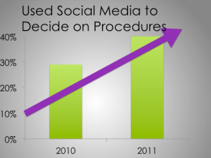
The American Society of Plastic Surgeons conducted a study looking at patients and the influence of social media regarding decisions on surgical procedures. In 2010, 29 percent of patients utilized social media that figure rose to 42 percent in 2011 and the numbers continue to increase.

Screen Shot 2014-08-24 at 3.20.07 PM


We are beginning to see websites like Vitals where patients can find doctors or facilities and write reviews. Vitals is a doctor rating site. They are trying to communicate that they are a trusted source. This is a great opportunity for us as physicians. We can’t necessarily operate on a site like Vitals, but we can on a website such as Health Tap. Health Tap is a question and answer site, i.e., people ask questions and experts answer them. When you answer the question, you get stars next to your name. 50,000 doctors went online to answer over a billion questions. This is an opportunity to demonstrate to patients that we are a trusted source. We are able to not only market to patients, but we can deliver something of value to them. These sites are very fast growing, particularly among providers.

RealSelf is another website similar to Health Tap; however, it is specifically for cosmetic surgery. RealSelf has 36 million unique visitors and there are 500,000 answers from board-certified physicians and healthcare professionals. Two hundred and thirty nine million dollars were spent by Real Self users on cosmetic surgery. This is a community that connects eager patients with trusted, vetted physicians. Their goal is to educate patients so that they can make informed decisions about cosmetic procedures. They post pricing on the website as well. This is an important trend that patients want to know about it as patients want transparency.

Eighty percent of people go online to search for health information. What do they see? If you Google yourself, you may find that you’re competing with restaurants and other reviews. “Is she or he a good doctor?” is a difficult question to answer. This is often substituted with “do I like him/her?”  If you think that review sites are going away, that’s not true. This is a good thing. If you’re doing the best thing(s) for your patients, these websites are going to help you. These websites work and patients get something out of them. You need to demonstrate this to your patients.

What can I do on my review site(s)?

Post a picture of yourself so that your patients can see you. Make sure that the information about your office/practice is accurate and say something personal about yourself. Patients are looking for personal information that connects you with them. This is free points for you and patients really care about it. You want to convey that you and your practice care about your patients. People want the “Apple” store experience when they come to see you.

What about Pinterest? Pinterest, if you didn’t know, is a tool that consumers use in order to find information on ideas and projects. There are 70 million users and 80 percent are females. This is important for us because 80 percent of women are the primary healthcare decision-makers among families. This is a great place to interact with women who are shopping. This is what women are interested in and it is an ideal place to showcase services. Harvard Business School refers to this as “reverse showrooming.” Reverse showrooming refers to when people browse online then purchase in a store. Pinterest is a big driver on in-store sales. 21 percent of users said they bought an item in-store after pinning, repinning, or liking it, and 36 percent of users under 35 said they had done so.

What about Facebook? What do people do on Facebook? Facebook is about feeling something. People go online to share pictures and talk about their families. If you go on Facebook, you have to be very careful about advertising. What about Twitter? Twitter is about now and relevance. What about YouTube? YouTube is a great place to convey information in an efficient manner. You can deliver value about a service. “If there’s anything more annoying than people talking about you, it is to have no one talk about you.” Oscar Wilde

Remember that social media is just one type of marketing. You should be using it along with traditional media and marketing tools. If you think social media is important now, wait until the next generation.


Melanoma Clinic: Lunch, Lesions, and Lessions

Part 1

Hensin Tsao, MD, PhD

Keith Flaherty, MD

Ilona Frieden

Philip LeBoit, MD

Dr Hensin Tsao, of Harvard Medical School, discusses several challenging cases with regards to the management of melanoma and pigmented lesions…Remember that melanoma, as an area, is not always easy.

Case 1

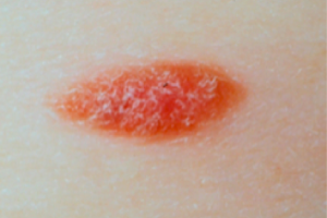
A ten year-old girl presents with a pink plaque on the left shoulder. The lesion has been present for four months and she has no other medical problems.

Screen Shot 2014-08-24 at 3.10.15 PM

As a clinician, what would you do next?

  1. Photo and follow up in 6 months
  2. Reassure and continue careful surveillance at home
  3. Perform an excisional biopsy with local anesthesia
  4. Biopsy under general anesthesia

Why not photograph and follow or reassure and continue surveillance. Because of the shape of this lesion, it’s a little bit bigger than normal spitz nevus. Because of her age, she can probably tolerate anesthesia so it would be worth obtaining a good specimen. The biopsy showed 1.5mm thick, Clark level IV, it was non-ulcerated and 2 mits/mm2.

Managing Pediatric Melanoma

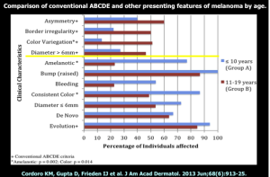
A study by Cordoro and colleagues demonstrated that pediatric melanoma does not follow the ABCDs. What do we look for? Most teenagers who get melanomas get adult-type melanoma. Up until age 13, melanoma is diagnosed at a relatively low level. Amelanotic or bleeding is also more frequent presentations of melanoma in younger kids.

Screen Shot 2014-08-24 at 3.10.28 PM

ABCDE Modified for Children

  • Amelanotic
  • Bleeding; bumps
  • Color uniformity
  • De Novo; Diameter; any
  • Evolution

Make sure that you have a dermatopathologist with whom you are very confident!

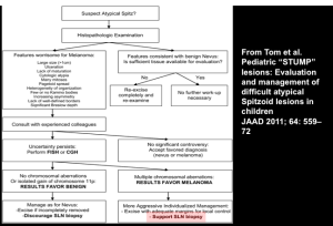
Risk stratification, rather than black and white diagnosis, is the way things are going now with regards to tumors and general pathology. There are lesions where we cannot always make a precise diagnosis, even after molecular testing. In many of these cases, all we can say is that it is a Spitzoid neoplasm with very low or high malignant potential. Spitzoid neoplasms can be stratified into four categories. These are Spitz nevus, Spitz nevus with atypical features, atypical Spitz tumor and Spitzoid melanoma.  Bastian and his colleagues have developed Spitzoid taxons:

  • H-ras mutated
  • Bap-1 mutated
  • Braf fusion
  • Alk fusion
  • Ntrak1 and ntrak3 fusion
  • Ros-1 fusion

One very interesting concept is that many of these genes, if you take them and translocate them in cells other than melanocytes, they are extremely potent in terms of oncogenic effect. ROS-1 and Alk, for example, are important players in lung cancer. Mutations or fusion partners do not define benign versus malignant, but only lineage. So, doing a test for Alk will not tell us if the lesion is benign or malignant; however, if the lesion does metastasize these are very important venues for therapy. For instance, BRAF, even though there’s no Braf mutation in these lesions, it is over-expressed and anti-BRAF therapies may be effective in Spitzoid melanomas that have a BRAF fusion. You don’t have to have the mutation for BRAF inhibition to work, if it’s a tumor where other things are turning it on.

Screen Shot 2014-08-24 at 3.10.49 PM


Zappin the Tats

Suzanne Kilmer, MD

At MauiDerm 2014, Dr Kilmer, an expert in lasers and light devices for dermatological procedures, discusses tattoo removal…

For tattoo removal, we generally use the visible spectrum of light.  Years ago, we used CO2 laser or salabrasion for tattoo removal. The use of the CW laser resulted in trans-epidemal loss of ink, significant thermal damage and frequent scarring. The newer Q-Switched Lasers shatters the ink into smaller particles then utilizes macrophages to remove the ink; there is some trans-epidermal loss, yet, there is rarely any scarring.

There are several issues to assess regarding tattoo removal. Pulse duration can be performed in either the nanosecond domain or more recently, the picosecond domain; the picosecond domain may be better for breaking up the ink and it is and may not be as wavelength dependent. Remember that longer wavelengths penetrate deeper and, very importantly, is the fact that the ink’s absorption determines the best wavelength. Fluence/spot size should also be considered. We want the largest spot size; however, we need sufficient fluence. Larger spot sizes allow deeper penetration of effective fluence as long as the laser has sufficient power. Regarding treatment intervals, six to eight weeks is generally the best timing. Of note, dark tattoos are much more easy to treat versus multiple colored tattoos. Green ink responds best to red light and red ink absorbs more in the green light.

Some of the side effects with tattoo removal include ink darkening, incomplete removal of ink, allergic reactions and infections.

Tattoo Treatment Improvements

Over 25 years ago, we went from long pulse to QS laser treatment. QS treatment demonstrated a dramatic improvement in ink reduction and side effect profile. This was the first time that we were able to remove tattoos without scarring. We started with 694nm then added 1064/532 and then 755nm wavelengths in order to improve color removal. This was performed for over 20 years with minimal improvement. In the last two years, we have shortened the pulse width to long pico/ultrashort nanosecond domain. Some studies demonstrated that multiple treatments in the same day increased ink clearance.

Picosecond lasers have a greater photoacoustic effect. In 2012, Cynosure came out with the first picosecond laser and it clearly demonstrated an increased clearance of ink when compared to the QS laser.

Picosecond lasers can also improve ink clearance in resistant tattoos with just two treatments when compared to QS lasers which showed no change over seven treatments.

Another company, Cutera, is looking at using other wavelengths with a novel picosecond laser. They are looking at a 1064nm with 532 component for picosecond domain.

Kossida & Anderson first reported faster tattoo clearing with single versus four treatments on the same day. After treatment, you should wait 20 minutes for whitening to clear “R20”. The photoacoustic effect and shattering of ink particles creates a cavity and as nitrogen gas flows in, you can see whitening. Nitrogen is slow to dissolve out (hence prolonged whitening). A study by Kilmer and Ibrahimi supported repetitive treatment on the same day in order to expedite tattoo clearance. They explored effectiveness of one versus two versus three versus four treatments (to alleviate scheduling nightmares.)  They found that two treatments were better than one but then they saw a declining efficacy with further treatment. Geronemus et al used an optical clearing agent, PFD, eliminating the 20-minute interval to clear whitening.

R20 Results-QS Laser

We have found that multiple treatments are better than single treatment on the same day for most tattoos. There is less of a difference between two, three and four treatments on the same day. Increased swelling may occur, as noted by patients and the treating nurse. There was no increase in pain; in fact, most patients felt less pain with subsequent treatment. We also saw no difference in PIPA and no scarring was noted. Generally, all patients preferred a more rapid tattoo clearance.

In summary, ultrashort pulse widths are better, but not always and multi pass is better, usually.


MauiDerm News Editor-Judy Seraphine


Rosacea

Richard Gallo, MD, PhD

In this presentation at MauiDerm 2014, Dr Richard Gallo, an expert in the field, provides us with an update on the management of rosacea…

Remember that rosacea almost exclusively affects the face, making it both a cosmetic and medical issue for those it affects. For the clinician, rosacea is a prevalent, serious, recurring and progressive dermatological condition that requires chronic therapy to prevent long-term sequelae.  Rosacea affects approximately 14 million Americans and possibly more. Unfortunately, the condition is often times not recognized; however, we have found that early recognition and treatment are important in order to prevent progression and disfigurement.  Therapeutic regimens must be tailored to the individual patients and need to address acute symptoms, help patients remain asymptomatic, and minimize the recurrence of symptoms.

Rosacea can be categorized into four sub-types: subtype 1-erythematotelangiectatic; subtype 2-papulopustular; subtype 3-phymatous; and subtype 4-ocular. (Wilkin J, et al. J Am Acad Derm 2002;46:584-587; Wilkin J, et al. J Am Acad Derm. 2004;50:907-912.) There are also many different variants of this disorder that do not always clearly fit into the above-mentioned subtypes such as vascular, sebaceous hyperplasia/rhinophyma and inflammatory causes.

The diagnosis of rosacea is based upon consensus opinions. We don’t have a clinical test or laboratory value, i.e., some sort of objective measurement that can determine rosacea.

Primary Diagnostic Features of Rosacea

  • Central facial erythema (fixed)
  • Flushing
  • Telangiectasias
  • +/- inflammatory lesions

Secondary Diagnostic Features

  • Burning or stinging
  • Plaque
  • Dry appearance and scale
  • Edema
  • Ocular manifestations
  • Peripheral location
  • Phymatous changes

(Del Rosso, J.Q., Thiboutot, D., Gallo, R.L. et al. Consensus Recommendations from the American Acne & Rosacea Society on the Management of Rosacea, Part 1. Cutis 2013; 234-240;

Wilkin, J, Dahl, M, Detmar, M, et al. Standard classification of rosacea: Report of the National Rosacea Society Expert Committee on the Classification and Staging of Rosacea. J Am Acad Dermatol 2002; 46:584.)

Unfortunately, rosacea is often misdiagnosed. Rosacea is variable in that there are various combinations of cutaneous signs, such as flushing, erythema, telangiectasia, edema, papules, pustules, ocular lesions and rhinophyma. Diagnostic confusion can occur because most patients only have some, rather than all, of these signs and symptoms. Other dermatological disorders and co-morbid conditions affecting the face can also cause confusion. Rosacea is often confused with acne and it may coincide with actinic keratosis, acne, seborrheic dermatitis, and contact dermatitis.

Inflammation is a major culprit! Inflammation in rosacea may result in capillary cell wall weakening secondary to dermal-matrix deterioration. It may also result in lymphatic failure secondary to elastin degradation and/or the production of vascular mediators. Inflammation precipitates episodic signs and symptoms of rosacea.

In order to really understand this disease, we need to understand the origin of inflammation. To come up with a way to effectively treat our patients we need to understand what causes this disease.

There are several environmental exposures that can trigger rosacea:

(Patient reported survey in rank order)

  •             Sun exposure
  •             Emotional stress
  •             Hot weather
  •             Wind
  •             Heavy exercise
  •             Alcohol consumption
  •             Hot baths
  •             Cold weather
  •             Spicy foods
  •             Humidity
  •             Indoor heat
  •             Certain skin-care products
  •             Heated beverages
  •             Certain cosmetics
  •             Medications
  •             Medical conditions
  •             Certain fruits
  •             Marinated meats
  •             Certain vegetables
  •             Dairy products

Can all of the subtypes and all of the different triggers really be the same disease?

Normal facial skin uses pattern recognition molecules to detect danger. Toll-like receptors are just one of many types of molecules that exist on the surface of keratinocytes and every cell type of the skin. Their goal is to detect patterns of the environment around them. Multiple microbes, dry skin and UV are all triggers of the pattern recognition system. Researchers began looking at biopsies of rosacea patients on non-inflamed skin. One particular concept was that rosacea patients have too much PRR (pattern recognition receptor) activity as seen by TLR2 protein staining.  This is an important issue to understand as it explains the essential role of general skin care as the skin has an excess of the molecular sensors of damage. UV injury as well as excessive dryness and even excessive oilyness triggers PRRs. This idea helps to put other theories into context.

Theories About Triggers

O’Reilly and colleagues published a paper demonstrating the positive correlation between serum immunoreactivity to Demodex-associated Bacillus proteins and erythematotelaniectatic rosacea. Eighty percent (21/26) of patients with erythematotelangiectatic rosacea showed serum reactivity to B. oleronius, compared with forty percent (9/2) of controls. (O’Reilly N, et al. Br J Dermatol. 2012) However, there are problems with this hypothesis. First of all this is fairly rare, one mite out of forty contained the bug and cross-reactivity of the bug antigens with other more common bugs has not yet been investigated. Many normal patients have this bug and did not react to it. Lastly, trying to kill Demodex is not usually effective for patients with rosacea. The take home message for current trigger data is that we, as clinicians, need to care for the barrier and remember that there is probably no single trigger for everyone. This makes patient history, clinical exam and your relationship with patients incredibly important.

What do the triggers do?

LL-37 expression is increased in rosacea. LL-37 is a very important molecule in a number of skin diseases in that it kills microbes as well as increases vasculature and inflammation upon its release. The other part of the equation that is important is the molecule KLK5, which is an enzyme that activates LL-37. So when we think about rosacea, remember that there are various signals that are activated by the environmental triggers.  This is variable based on genetic makeup and the ecological organisms on the skin. A normal innate immune system reacts to infection and true danger, there is no inflammation in a resting state. Rosacea patients; however, have a molecular sensitive skin because of too much PRR. Once that is triggered, the trigger system makes too much KLK5 that can activate LL-37 and initiate certain aspects of this disease.

It is important to understand the role of KLK and LL37 in rosacea because they explain the benefits of current therapies. They may also serve as a potential objective biomarker as well as aiding in future new drug development. Dr Gallo and many others have worked in larger studies to validate and confirm these observations.  A blinded study conducted by Coda and colleagues looked at the KLK enzyme on the skin and found that rosacea patients have elevated enzyme activity but divide into two populations that weren’t necessarily distinguishable on clinical presentation. This may be important because protease level can predict topical azeleic acid response. Patients with high enzyme activity who were treated with azeleic acid showed a significant drop in activity over sixteen weeks, whereas patients who started off with low protease activity showed no response. (Coda, et al. JAAD. 2013)

Other therapies that we currently have may also be working because of a similar process. The tetracycline class drugs have shown to inhibit this enzyme. When we look back at old literature, we see that some of the biochemists found that one of the things that tetracycline seemed to do was inhibit enzyme activity completely independent of any action on a bacteria.

Testing the Hypothesis: Does targeted inhibition of KLK5 activity improve rosacea?

Dr Gallo and colleagues conducted a pilot clinical trial using an FDA-approved serine protease inhibitor that was formulated into a topical cream. It was a 12-week, double-blind, placebo controlled BID application to measure both protease and clinical response. The only patients who improved in this trial were those who applied the protease inhibitor. (Two, A.M. et al. J Invest Dermatol. 2013) This is not necessarily proof of a new therapy, but it is intriguing data that may prove the theory relevant.

In summary, rosacea patients have high KLK5 and LL37. KLK5 activates cathelicidin and more LL-37 and KLK5 is associated with disease severity. Successful therapies are associated with decreased KLK5 activity or cathelicidin.

Neuromediators Are Important!

Neuromediators increase vasodilatation, nerve endings, neuropeptides and they stimulate mast cells. What can we do about this? We have alternative neuromediators that fight these vasodilitation effects. The alpha-adrenoceptor agonists are the stimuli are the alpha1 and alpha2 receptors. They have the ability to initiate smooth muscle contraction in a dilated vessel. When topically applied, both brimonidine (a2 agonist) and oxymetazoline (a1 agonist and a2 agonist) have shown to be successful therapies for the erythematous stage of rosacea. (Fowler J et al. J Drugs Dermatol. 2013; Shanler and Ondo Arch Derm 2007)  These are alternative mechanisms thinking about the pathology of the disease. The drivers of the disease are the molecules that initiate inflammation and vascular growth. We now have strategies for inhibiting or reducing LL-37 and limiting vasoactive response.

Topical Rosacea Therapy

The goal of topical therapy is barrier repair, decrease triggers, decrease LL-37 and vasoconstriction. Don’t forget to always include appropriate barrier care such as sunscreen, gentle cleansers and moisturizers.

  • Azeleic acid
  • Sodium sulfacetamide/sulfur
  • Brimonidine
  • Metronidazole
  • Tretinoin
  • Tacrolimus/pimecrolimus
  • BP/Clinda
  • Ivermectin

Oral Rosacea Therapy

Oral therapy targets triggers and drivers.

  • Tetra/doxy/mino
  • Cipro
  • Bactrim
  • Isotretinoin

Clinical Pearls

Early Rosacea

  1. Trying to minimize triggers
  2. Rosacea drivers not major yet
  3. Complaint is erythema

What should we do?

  1. Trigger avoidance
  2. Barrier care
  3. Topical agent
  4. Adrenergic agonist

Moderate/severe Papulopustular

  1. Still need to minimize triggers
  2. Rosacea drivers active
  3. Complaint is red papules

What should we do?

  1. Oral agent (TCN)
  2. Barrier care ±sunscreen
  3. Topical agent
  4. Adrenergic agonist
  5. Second line Isotretinoin

Perioral Rosacea

  1. Spared vermillion
  2. Microbial influence?
  3. Ask about steroid

What should we do?

  1. Oral agent (TCN)
  2. Barrier care
  3. Possible TIM

In conclusion, we are finally getting a mechanistic understanding of rosacea. There is a need for an individual approach and both rosacea triggers and drivers need to be identified and targeted. We have good and complementary options for medical therapy. Do not forget the importance of barrier care!!


MauiDerm News Editor-Judy Seraphine

Systemic Medications: Top Ten Take Home Points

Matthew J. Zirwas, MD 

  1. Prednisone is drug of choice for expected short term therapy – weeks.
  2. Cyclosporine is drug of choice for expected “medium term therapy” – months.
  3. Methotrexate is drug of choice for chronic therapy in patients without liver, kidney, or hematologic co-morbidities.
  4. Mycophenolate is drug of choice for chronic therapy in patients with liver, kidney, or hematologic co-morbidities.
  5. Cyclosporine has many drug interactions – must use a computerized drug interaction checker.
  6. Methotrexate has a few drug interactions but they are extremely serious and can be fatal.
  7. Mycophenolate has few drug interactions.
  8. Patients on methotrexate should take folic acid on days they do not take the methotrexate.
  9. Older patients on mycophenolate, in particular, are at high risk of zoster.
  10.  If patients on cyclosporine have elevations in creatinine, they need to stop the medication.

Pigmented Lesions: Clinical Pearls

Ashfaq Marghoob, MD

Dr Marghoob provides us with his clinical pearls on pigmented lesions….

  1. The larger the congenital nevus the greater is the risk for developing melanoma.
  2. The risk of a melanoma developing in a small congenital nevi is small enough that prophylactic excision is not required.
  3. Presence of many nevi and dysplastic nevi are strong risk factors for melanoma.
  4. Analytical, Differential and comparative recognition are all helpful in differentiating nevi from melanoma.
  5. Total body photography assists clinicians in finding concerning lesions.
  6. Dermoscopy assists clinicians in deciding which lesions require a biopsy.
  7. Unna’s theory of nevogenesis is not supported by recent cross sectional and longitudinal studies.
  8. Nevi with a peripheral globular pattern or a starburst pattern are growing nevi (not yet in senescence).
  9. Most halo nevi have a globular pattern.
  10. Patient driven healthcare with the use of Apps is likely to help in finding early melanomas.

Immunology 101: The Basics

Andrew Blauvelt, MD

Dr Blauvelt provides us with the 10 most important take-home messages from his immunology presentation at MauiDerm NP+PA Summer 2014….

  1. Key features of the innate immune system include: rapid response, non-specificity, phagocytosis, no memory.
  2. Key features of the  acquired immune system include: slow response, very specific, lymphocyte-mediated, memory.
  3. Keratinocytes are active participants in generating immune responses by secreting numerous cytokines upon activation.
  4. 4.Toll-like receptors are pattern recognition receptors on keratinocytes that recognize foreign antigenic material.
  5. Antimicrobial peptides are natural antibiotic molecules found in skin that are abundant in psoriasis skin and sparse in atopic dermatitis skin.
  6. 6.Epidermal Langerhans cells are antigen presenting cells that recognize/process skin antigens and migrate to lymph nodes, where they present antigen to T cells.
  7. 7.T cells require 3 signals to become fully activated: 1) recognition of antigen by the T cell receptor via MHC on the surface of antigen presenting cells; 2) binding of co-stimulatory molecules on T cells and antigen presenting cells to one another; and 3) secretion of soluble cytokines by the T cells.
  8. CD4+ T cells are T helper cells that recognize antigen via MHC class II and secrete cytokines to enhance CD8+ T cell and B cell immune responses, whereas CD8+ T cells are cytotoxic T cells that recognize antigen via MHC class I and kill cells upon contact.
  9. B cells secrete antibodies that specifically bind to antigen.
  10. Primary immune responses are slow, occur after first exposure to antigen, and involve creation of memory cells, whereas secondary immune responsesare fast, occur after subsequent exposures to antigens, and involve reactivation of memory cells.


Dermatitis Overview: 2014

Matthew J. Zirwas, MD

Are you treating dermatitis? Dr Zirwas provides us with ten clinical pearls…

  1. The main goal in dermatitis patients is figuring out WHY they have dermatitis.
  2. Acute allergic contact dermatitis starts 1-3 days after exposure to the allergen and takes 1-3 weeks to go away.
  3. Chronic allergic contact dermatitis takes 3-6 months to go away.
  4. Hypoallergenic rubber gloves are not hypoallergenic.  Hypoallergenic gloves include: vinyl gloves, Microtouch Nitra-Free, Dermaprene Ultra.
  5. Shampoo and conditioner are the main causes of eyelid dermatitis, both irritant and allergic.
  6. Atopic dermatitis is the combination of inadequate skin barrier and an immune system that was genetically primed to go in an allergic direction.
  7. Food avoidance and chelation do not work in adults.
  8. There are probiotics that work, but it is very specific – patients can’t just take any old probiotic.
  9. Seborrheic dermatitis is an inflammatory reaction to Malassezia yeast on the skin – treat by reducing amount of yeast and targeting the inflammation.
  10. Xerosis is too little water in the skin.  Need to add water and prevent it from just evaporating away.