Dermoscopy: Clinical Pearls

Ashfaq Marghoob, MD

Are you performing dermsocopy in your clinical practice? Dr Marghoob provides us with his clinical pearls…

  1. Dermoscopy helps in differentiating benign lesions from skin cancer.
  2. The presence of network (reticular network, negative network, polygonal lines), streaks (radial streaming, pseudopods), aggregated globules/peripheral globules, homogeneous blue pigmentation is most often seen in melanocytic lesions.
  3. Melanocytic lesions that deviate from the 10 benign patterns and has at least one of the 10 melanoma-specific structures needs to be biopsied to rule out melanoma.
  4. The presence of spoke wheel structures and/or leaf like areas is 100% specific for BCC.
  5. Comedo like openings and milia cyst are often seen in SK but can also be seen in other lesions including melanoma.
  6. Polarized light makes blood vessels and crystalline structures more conspicious, and makes milia cyst less conspicuous.
  7.  A polymorphous vascular pattern in an amelanotic lesion should raise concern for melanoma.
  8. Dermoscopy improves the clinician’s diagnostic accuracy.
  9. All structureless or featureless or not-otherwise-diagnosable lesions should be viewed with suspicion.
  10. Raised lesions should never be monitored for change.

Cutaneous Oncology: Talking Points

Marc Brown, MD

Here are some of the key points from Dr Brown’s presentation at MauiDerm NP+PA Summer 2014….

  1. High risk parameters for BCC include location on the central face, larger size, recurrence, prior radiation and aggressive histology.


  1.  High risk histology for BCC includes the following:  infiltrating, morpheaform, micronodular, basosquamous, sclerosing, desmoplastic, and perineural invasion.


  1. Risk factors for the development of SCC include:  UV light exposure, X ray exposure, HVP infection, immunosuppression, chronic non-healing or inflammatory wounds, an rare genetic syndromes.


  1. High risk locations for SCC are ear, lip, genitalia and scalp.


  1. Other risk factors for aggressive SCC include:  poorly differentiated histology, depth of invasion, perineural invasion, size (greater than 2 cm.).


Emerging Therapies in Psoriasis: Clinical Pearls

Bruce Strober, MD, PhD

What’s new in the field of psoriasis? Dr Strober provides us with some clinical pearls…

  1. TNF-inhibitors cause weight gain.
  2. Ustekinumab does not cause weight gain.
  3. IL-23 inhibitors block p19 and are more specific than IL-12/23 inhibitors, which block p40.
  4. Apremilast achieves PASI75 in approximately 30% of patients after 16 weeks.
  5. IL-17 pathway inhibitors achieve PASI75 in approximately 80% of patients after 12-16 weeks.
  6. IL-17 pathway inhibitors may slightly increase the risk of mucocutaneous candidiasis.
  7. JAK kinase inhibitors will require monitoring for liver function, renal function, lipids and creatinine phosphokinase.
  8. JAK kinase inhibitors might increase the risk of varicella zoster infection.
  9. Apremilast treats psoriatic arthritis.
  10. Apremilast does not need laboratory monitoring.


10 Pearls from Dermatopathology – The Biopsy, Analysis & Report

Whitney J. High, MD, JD, MEng

  • Dermatopathology is one of  two ABMS-recognized subspecialties in dermatology, and one may become fellowship trained after first being a board-certified dermatogist or general pathologist.
  • Biopsy use is increasing.  In nine geographic areas of the USA, over the time period 1986-2001, the biopsy rate among those >65 years of age rose 2.5-fold, and the melanoma rate rose 2.4-fold.
  • There are multiple steps involved in taking a specimen from a piece of “wet” tissue, in formalin, to an interpretable slide and to a typewritten report.  These steps include:

Screen Shot 2014-06-25 at 9.14.06 AM

  • The dermatopathologist is examining but a small portion of your original sampling, and    this must always be considered when one assesses the “representative nature” of the results.
  • There is an old mantra in pathology in general: crap in = crap out.  No dermatopathologist, regardless of skill or expertise, can weave a poor sampling into an outstanding result.
  • It is the clinician responsibility to secure a “representative biopsy”, and if this is not done, eventually, this inadequacy  will be discovered.  Over the period of 1998-2005 the number of shaves increased but the volume of shaves decreased.
  • The technique employed (shave, punch, excision) must be adapted to the clinical situation – there are no fixed rules that may be applied to every situation.  This is why the clinician is being paid an “evaluation/management” code; namely, to select a biopsy that is appropriate for the circumstances.
  • A recent study of pigmented lesions showed the odds of misdiagnosis (overall and associated with an adverse outcome) were much higher with a punch biopsy than with an excisional biopsy, whereas a shave biopsy was only weakly associated with misdiagnosis. (Ng et al. 2010)
  • Situations where the biopsy technique should be carefully considered include suspected:

Screen Shot 2014-06-25 at 9.16.42 AM

  • The pathology report itself should be carefully read and scrutinized to understand precisely what the dermatopathologist is trying to convey. Demographic data should be confirmed. The technique and specimen size should be verified. Data used by the dermatopathologist to formulate the diagnosis should be noted  (i.e., step levels, immunostains, special stains, etc.). If questions still exist, a phone call should be placed to the dermatopathologist for expanded dialog.

Eruption! Pediatric Acne

Larry Eichenfield, MD

Dr Eichenfield provided the audience at MauiDerm 2014 with an update on the new pediatric guidelines for the management of acne. These guidelines, published in May of 2013, were developed by the American Acne and Roseacea Society and endorsed by the American Academy of Pediatrics, which is the first time that the Academy has a set of guidelines for the management of acne. One of the main reasons this was done was to address the practice gap between dermatologists and pediatricians. The group of experts was comprised of dermatologists with expertise in acne, pediatric dermatologists, and pediatricians.

As we know, acne ranges in terms of presentation and severity.  Acne can be categorized by age, i.e., neonatal (0-6wk), infantile (0-1), mid-childhood (1-7), preadolescent (7-12), and adolescent (12-19).

Dr Eichenfield emphasizes that mid-childhood acne (age 2 – 7) acne is the most worrisome. It can be associated with premature adrenarche, Cushings Syndrome, CAH, gonadal/adrenal tumors, and precocious puberty. Mid-childhood acne is very uncommon, and is a sign of early adrenarche, often associated with a pathologic process. The assessment for mid-childhood acne includes assessment of  testicular size (males), presence or absence of hirsutism, clitoromegaly, androgenetic alopecia, increased muscle mass, and/or deepening of the voice (males).  Tests and examinations include a growth chart, evaluation including bone age, tanner stage, Total/Free testosterone, DHEAS, androstenedione, LH, FSH, prolactin, and 17OH-progesterone. The guidelines suggest that you should refer these patients to a pediatric endocrinologist.

What do the new guidelines say?

It’s important to know that pre-adolescent (7 ≤ 12 years) acne is common and may precede other signs of pubertal maturation. Work-up beyond history and physical is generally unnecessary unless there are signs of androgen excess, polycystic ovarian syndrome, or other systemic abnormalities. According to the data, there is evidence that acne, and possibly puberty, are now occurring at an increasingly earlier age for both males and females. If you look at some of the papers that were published about a decade ago, the amount of comedones that were seen in a ten or 12 year-old then is probably different now. Twelve is no longer an age point defining “normal acne;”  if you are eight and above acne can be typical and common.

After the guidelines were published early acne in preteens was highlighted in several newspaper articles, and radio and television segments.  Articles were published in both the New York Times and USA Today. In fact, after the New York Times article was published, there were over 157 blogs that provided an interesting perspective comparing what the general public thinks regarding acne and what we do as specialists.  A recent analysis of the National Ambulatory Medical Care Survey (NAMCS) database assessed trends in the age of children seeking treatment for acne. NAMCS data from 1979 through 2007 were analyzed for all physician visits in which acne vulgaris was listed as a diagnosis in children aged 6 to 18. The analysis revealed a significant decrease in the mean age of children seeking treatment for acne over this period.

Over the past several years, clinicians have noted an earlier onset of acne—that is, the appearance of acne in patients as young as 8 or 9 years. Indeed, it has been suggested that 12 years of age should no longer be considered the low end of the “normal” range for the onset of acne.

Acne may be the first sign of the onset of puberty in children aged 7 to 11 years. The general clinical impression of earlier puberty is, in fact, supported by epidemiologic data in the US and elsewhere. In general, the trend toward earlier puberty is stronger among girls than among boys. In any event, the earlier onset of acne has mirrored the downward trend in puberty timing.

The guidelines provide an algorithm for the management of pediatric acne. If you can generally categorize the acne as mild, moderate or severe, you can access the algorithm. Keep in mind that this algorithm is slightly different than prior guidelines, in that for mild acne initial treatment benzoyl peroxide, based upon the evidence, made it as one potential, initial solo therapy.   The guidelines can be found in the Journal of the American Academy of Pediatrics, or online at: http://pediatrics.aappublications.org/content/131/Supplement_3/S163.full.pdf.

Acne Guidelines: Highlights

The guidelines emphasize appropriate use of medications based upon disease severity.  Oral antibiotics should be used concurrently with a topical retinoid because it is important to build a topical regiment to “transfer the patient to” after a limited course of antibiotics.   A variety of studies show that 70 percent of the time you can transition your patient who is on an oral antibiotic and topical retinoid to a regiment of topical retinoid alone or combinations with topical antimicrobials (like benzoyl peroxide) and/or antibiotics

Topical retinoids may be used as monotherapy or in combination products and in regimens of care for all types and severities of acne in children and adolescents of all ages. It is important to remember that topical antibiotics are not recommended as monotherapy and if a topical antibiotic treatment is to be used for more than a few weeks, topical benzoyl peroxide should be added, or used in combination products. The guidelines also suggest that fixed-dose combination topical therapies may be useful in regimens of care for all types and severities of acne.

With regards to oral antibiotics, they are a reasonable approach for moderate to severe inflammatory acne vulgaris at any age. Tetracycline derivatives should not bee utilized in children 8 years of age and below. Second generation tetracyclines are “sometimes preferred” to tetracycline because of ease of use, fewer problems with absorption with food and minerals in vitamins and other supplements, and less frequent dosing. It’s very important that patients are educated and monitored for potential adverse events when utilizing oral antibiotics for acne.

Hormonal therapy is actually interesting because there are people who are very pro-hormonal therapy and others are a bit more conservative and prefer oral antibiotics. The group ended up stating that combined oral contraceptives (OCs) may also be useful as second-line therapy in regimens of care in pubertal females with moderate to severe acne; however, tobacco use and family history of thrombotic events should be assessed. Due to concerns about growth and bone density, many recommend withholding OCs for acne until one year after onset of menstruation.

Isotretinoin is recommended for severe, scarring, and/or refractory acne in adolescents and may be utilized in younger patients. Remember that extensive counseling, particularly regarding the avoidance of pregnancy, as well as careful monitoring of potential side effects and toxicities, is recommended. As far as the specific discussion on isotretinoin in pediatric use, there are bone mineralization changes; however, the data is inconsistent and it is not associated with increased factures. Hyperostoses are very uncommon for acne and there has been one case of premature epiphyseal closure in a patient on isotretinoin for acne, but that’s not necessarily attributable to the isotretinoin. IBD is controversial, but counseling is reasonable.

Practice Gaps

Dr Eichenfield states that there is a chasm in the way that General Practitioners, Pediatricians, and Dermatologists treat acne. A 2014 paper by Tan et al showed that topical retinoids were prescribed in 41 percent of acne visits. Older age, male gender and having Medicaid insurance were associated with a lower likelihood of receiving a topical retinoid prescription. Moreover, the researchers found that in the Medicaid dataset, patients who saw a pediatrician or general practitioner had lower odds of receiving a topical retinoid prescription versus those patients who saw a dermatologist.

Another study looked at the National Ambulatory Medical Care Survey (NAMCS) data regarding the treatment of preadolescent acne in the United States. The data were stratified according to age group and physician specialty. The findings are presented below:

Screen Shot 2014-06-11 at 1.39.02 PM


What are Dr Eichenfield’s thoughts on the practice gaps?

  • Over-reliance on oral antibiotics
  • Use of oral antibiotics without BP
  • Use of oral antibiotics without Retinoid
  • Use of topical and oral antibiotics together, without retinoid
  • Under appreciation of early, significant acne as predictor of worse acne over time

Literature has shown that early comedonal acne may predict later severe acne. So remember that if you see a patient with a high number of comedone counts at an early age, their chances of severe inflammatory acne at a later age is much higher than someone who has low comedone counts early on.

Another issue that Dr Eichenfield is appreciating more and more in clinical practice is that you can have very early, subtle scarring. We know that there is a lot of scarring that occurs without nodular-cystic disease, so this is very common. This is an important to try to get patients evaluated early so that scarring can be prevented, and minimized. Dr Eichenfield advised  that a useful technique is to side-light the face and look for depressions in the face, displaying  scarring as opposed to post-inflammatory hyperpigmentation or persistent erythema.

A study by Patel and colleagues aimed to determine what types of acne lesions preceded the development of atrophic acne scars. Twenty-two patients with mild to moderate acne were enrolled in a split-face study in which one side was treated with non-ablative laser and the other remained untreated. A series of standardized digital facial photographs was obtained from the untreated side at 2-week intervals from baseline to week 12, and all photographs in the series were aligned with the baseline photo. When all of the atrophic scars were tracked to baseline, 53 were found to have arisen from clinically normal skin, 30 were established scars, and 21 arose from acne lesions, including closed comedones. However, no open comedones at baseline corresponded to atrophic scars.  The results of this study not only verify that inflammatory acne lesions often lead to atrophic scarring, but also demonstrate that acne scars may arise from initially comedonal lesions, as well as from clinically normal skin. Moreover, they indicate that a period of 12 weeks is sufficiently long to develop and establish atrophic scars. Thus, aggressive treatment of both inflammatory and comedonal acne is warranted to minimize acne scarring.

What about Isotretinoin?

We know that isotretinoin is the most effective treatment for acne; however, the optimal dosing regimen is still unknown.  Dr Eichenfield comments that he uses isotretinoin commonly. He also states that he tends to be biased towards lower-dose isotretinoin on a daily basis, working up to cumulative doses of 120-150 mg/kg.  He commonly will the daily doeses up to the “highest comfortable dose,”  that is, the highest dose with minimal significant side effects or laboratory abnormalities.

A recent publication looked at 180 patients with acne resistant to other treatments who were enrolled in an observational, prospective study of istotretinoin with cumulative doses less than to 220 mg/kg versus isotretinoin greater than or equal to 220 mg/kg. Of these patients, 116 participated in the 12-month follow-up survey. At that time, 97.4 percent of the patients reported that their acne was improved. Overall, acne in 32.7 percent of the patients in the study relapsed at 12 months, and 1.72 percent of the patients required a retrial. In the lower-dose treatment group, the relapse rate was 47.4 percent compared with 26.9 percent in the high-dose group. Almost 100 percent of the patients in both treatment groups developed cheilitis and xerosis during treatment. Retinoid dermatitis was significantly more common in the high-dose treatment group and none of the other adverse effects were significantly different between the two groups.  However,  it should be noted that in the higher dose group,  nine patients had  persistent muscle aches, eight patients had persistent joint aches, and two patients  had hearing changes. (Blasiak RC et al. JAMA Dermatol 2013;149(12):1392-1398) Also of importance with regards to this study are the laboratory abnormalities based upon the dosing. (See table 1)

Screen Shot 2014-06-11 at 1.39.15 PM


Dr Eichenfield states that this publication has yet to “move him” to abandon his current methodology with regards to isotretinoin dosing for this patients.

Idiopathic Facial Asceptic Granuloma (IFAG) and Childhood Rosacea

We have seen a very interesting change in perspective regarding this disease. Occasionally, we see these patients who present with lumpy, cystic-type lesions, separate from acneiform lesions.   A multi-center study of four French dermatologic centers looked at patients who were diagnosed with IFAG between October 2000 and July 2007. Thirty-eight patients were included in the study. The median age at the time of diagnosis of IFAG was 43 months, with a median follow-up of 3.9 years. Sixteen patients (42.1%) had at least two criteria of childhood rosacea, 11 of 32 (34.4%) with a single lesion and 5 of 6 (83.3%) with multiple lesions. Remember that childhood rosacea presents with flushing, permanent or recurring erythema, papules and postules without comedones or microcysts, convexity predominance of lesions, ocular rosacea (chalazions, conjunctival hyperemia, keratitis). (Prey S, Ezzedine K et al. Pediatric Dermatology 2013;30:429-32)

What does this mean to us? Children with IFAG are at risk for childhood rosacea, and follow-up is advised, including periodic ophthalmologic assessment.


MauiDerm News Editor- Judy Seraphine


Hot Papers in Acne

Written by: Judy Seraphine

At MauiDerm 2014, Dr Webster lead off the discussion by reviewing some of the hot papers in acne in 2013 that help with either the understanding of acne or the treating of it. The first paper, published in JAMA Derm, discusses high dose isotretinoin and whether or not it is safe and effective. Traditionally, data demonstrated that the 120 mg/kg dose has a 20% relapse rate, this is based upon youth, severity, diet, and hormones. We have learned that if someone has really bad acne as a child, they will most likely have a relapse later and likewise, patients who had really severe acne on isotretinoin will likely relapse. A paper published in the International Journal of Dermatology in 2012 demonstrated that isotretinoin 290 mg/kg had a 12 percent relapse rate over three years; of note, adverse events were not any worse. Another retrospective paper, published in JAMA Derm in 2013, showed that isotretinoin greater than 220 mg/kg had a relapse of 27 percent versus 48 percent in doses less than 220 mg/kg—so the really high dose had a lower rate, but it was still somewhat high. Those who received the higher dose did have increased dermatitis.  Another finding from the study was that those on the higher dose of isotretinoin were aching a lot longer even when the isotretinoin was over; this is something that we don’t see with the standard dosing. As dermatologists, we have to consider that there may be a difference in how we define relapse and prospective head-to-head studies need to be done.

A paper, published by Drs Webster and Leyden in the JAAD, looked at a new form of isotretinoin that doesn’t require a dietary adjustment. We have known for years that isotretinoin requires a fatty meal, as there is decreased absorption on an empty stomach; in fact, there is a 50 percent decrease of isotretinoin if taken on an empty stomach. Dr Webster feels that the most common cause of isotretinoin resistance and relapse is due to patients taking the drug on an empty stomach. Data exist demonstrating that high-fat meals enhance the absorption of the drug, yet this new form of isotretinoin may be a viable alternative with regards to dietary change and acne outcomes. Dr Webster comments that whether or not you use this new drug or regular isotretinoin, it really should be taken with some fat in the stomach.

Figure 1 depicts the absorption rates with Accutane 40mg “fed” versus “fasted” and you can see that there is a significant difference.

Screen Shot 2014-05-26 at 8.06.28 AM

With the new product, isotretinoin-lidose, you can see that the absorption rate in Accutante (fasted) is exactly the same; however, the newer form of isotretinoin (fasted) is much higher. This drug may be helpful for compliance-challenged patients. (See Figure 2)

Screen Shot 2014-05-26 at 8.06.37 AM

What about P. acnes?

Recently, there have been a couple of papers that have been published suggesting that there may be a particularly hot strain of P. acnes that is causing acne. Years ago, one of Dr Webster’s first papers was looking at all of the strains of P. acnes with a bacteriophage typing system and they found that there were many strains of P. acnes that were reproducibly different, but there was no difference in where they showed up, i.e., the same spread of strains whether it was inflammatory or non-inflammatory acne or whether it was isolated from CNS surgery or from hip infections.

A recent paper, published in the Journal of Investigative Dermatology, reported that they found two strains of P. acnes. The type 1a strain of P. acnes predominates in inflammatory acne lesions and 1b in non-inflammatory; therefore, some strains are associated with inflammatory acne. A second paper, published in the British Journal of Dermatology, found that this association; however, is a trend and not an absolute, i.e., there were patients with bad acne who had the “non-inflammatory” strain and there were patients with no acne who had the “inflammatory” strain. Typically patients with inflammatory acne had a mixture of the two with the “bad strain” predominating.

How do you explain this and does it make sense? Does it mean that people with bad acne need to have their P. acnes replaced with a non-verulent strain so that they don’t get pimples? Or is this an ecological issue? The skin microbiome is a microecology in the same way that a swamp has its own ecology. Different selective pressures do different things to the microenvironment. For example, if you sample the forehead, it is very rich in P. acnes and malassezia. If you sample the forearm, there are usually no sebaceous glands and there is no food for P. acnes so you don’t get a lot of growth from P. acnes. This tells us that big variations in environments lead to big variations in strains.

Dr Webster suspects that this difference in P. acnes distribution between acne and non-acne is not necessarily “bad strain” versus “good strain”, it’s that inflammation is a selective pressure just like the presence or absence of food/water in an ecosystem; this adds oxidative stress on the bacteria. Remember that P. acnes defends itself against oxidation pretty well even though it is an anaerobe. Dr Webster feels that the enrichment of type 1a strains in patients with inflammatory acne is merely showing that inflammation selects through strains that can survive better in a more oxidized environment.  As dermatologists, we really need to look at how strain 1a is different from strain 1b—does it defend itself better against oxidation?


Introduction to Lasers and Light (And a Touch of RF and Microwave)

E.V. Ross, MD

Dr Ross, a leader in the field of laser and light therapy, provided the audience with an in-depth overview on lasers and light. Dr Ross begins by reminding us that we tend to look for the “easy” way out to make our patients look their best with the least downtime. In order to be efficient and effective for his patients, Dr Ross likes to have many lasers in one room; therefore, providing the best possible technology.

It is important that we really understand laser physics. You need to know some math, but it doesn’t have to be difficult math. You can take very complex mathematical relationships and break them down into very simple algebraic relationships. By doing so, you can actually apply your lasers and other technologies quite confidently. It’s imperative to understand how they work because if you don’t, you can get into trouble very fast. Remember to look, listen and feel….listen to the reaction, look at the laser, look at the patient at every pulse to be sure the endpoint is what you want to see because ultimately, the endpoint is more important than the physics.

When we talk about lasers, we need to know some basic definitions. One of the most important terms is fluence; fluence is simply the light dose, i.e., the amount of energy that we are investing per surface area for a particular application.  We used to speak a lot about power, e.g. 7 watts power or 12 watts power. Currently, we don’t talk much about power because most of our lasers today are pulsed lasers. What really matters are wavelength, fluence and pulse duration. Pulse Width is  very important, i.e., the time over which energy is delivered. Spot size, which contributes to the intensity inside the skin, is mainly important for visible light lasers and infrared lasers because a larger spot will penetrate better.

LASER, Light Amplification by the Stimulated Emission of Radiation, is a concept that Einstein predicted back in the 1920s, but it was not realized until May of 1960.

Why is laser different than a lot of other light sources? We can use a lot of non-laser light sources in dermatology and certainly achieve nice results. Intense pulsed light is a great example. There are some features of lasers that make them helpful for dermatology and helpful for certain applications, but not always necessary for every application.  (See http://www.colorado.edu/physics/2000/lasers/index.html)

Why is it important to differentiate laser light from non-laser light? Dr Ross explains that it is because of engineering more than anything else. Most of our targets that we treat in dermatology have multiple wavelengths as far as their absorption. Lasers are a convenient way to deliver light, i.e., deliver photons to the target. This is why lasers are so popular. You can put lasers into a fiber, you can have monochromatic light, and you can deliver very high power. It’s the only way to really deliver very short pulses in nanoseconds and picoseconds for certain applications.

Why do we need to know how/why lasers, etc. work?

Dr Ross states that one important reason is that when your laser breaks down, which can happen, you want to understand why it isn’t working. If you have a good understanding of how your laser works, you may be able to correctly diagnose that. If you see that your laser isn’t working right, often times there’s an error code. It’s important that you write down the code and report that to the technician. Sometimes it’s as simple as restarting the system.

You should be able to troubleshoot laser problems in a logical way…Device malfunction is one problem, an example is a temperature sensor malfunction. However, a bigger problem could be your lack of familiarity with the device; most laser problems are caused by the operator because they’re not familiar with how the device works—sometimes they are rented and sometimes they are only used once per week. Other factors that contribute to laser problems include poor patient selection, operational errors, poor post-treatment care, and simply bad luck.

Dr Ross reminds us that we have to think. Most of the time when we get into trouble with lasers it’s because we weren’t thinking—invest all of your brain power into that particular case while you’re performing the procedure and that means paying particular attention to endpoints.

Four years ago right before the Christmas holiday, Dr Ross had a typical case of a woman who came in for treatment of telangiectasias with a pulsed-dye laser. He used the pulsed-dye laser, 10mm spot, 7 J/cm2, and a 10-millisecond pulse and everything was looking fine. The patient developed some mild purpura and she was incredibly upset. So what happened? She didn’t have any blisters and everything else was fine. Dr Ross went back and looked at the beam profile and found that it was off by about 2mm (8mm instead of 10mm).  What had then happened was that the fiber was damaged so they were delivering the right energy but in a smaller spot; therefore, causing the fluence to become higher and the purpura threshold had become breached. The technicians then fixed the beam profile and there was no more purpura. Dr Ross reminds us that sometimes we may have to do some detective work to figure out what’s going on.

Broken mirrors can also compromise your laser experience.  Particularly with the CO2 laser, if your beam profile doesn’t look good it may be due to a broken mirror.

When your calibration doesn’t pass,  the most common reason is because of a bad lamp.

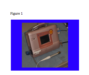
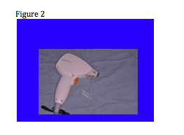
What we’re seeing now is a progressive change in laser technology. Lasers are becoming quieter, cooler, smaller, better and more reliable. We now have small, portable lasers (see figure 1) and maybe, in the near future, we’ll see small lasers that can treat everything from tattoos to resurfacing.

Screen Shot 2014-05-26 at 7.50.01 AM

Remember that with most of the lasers that we’re using today, the light piece is “back in the box” and is delivered through some sort of delivery system; however, in the case of some of the mini diode lasers, the light is in the hand piece. (See Figure 2)

Screen Shot 2014-05-26 at 7.50.10 AM

Over the next few years, we are also going to see more and more LEDs. We are going to begin to see more safety features that will be incorporated into the software with touchscreens. For example, you may have a patient who came in for treatment of a wart so the technician turns the dynamic cooling device off. Then, your next patient is coming for treatment of a port wine stain and the technician forgets to turn the DCD back on and the patient gets a blister from the very first pulse. What if there was a tool that provided a warning signal indicating that the DCD was off? Actually that feature is already built into one popular pulsed dye laser.

What about photon recycling? This is a way to capture some of the photons that were wasted. Whenever we use a laser much of the energy is reflected back off the skin surface. By recycling the energy we have a second chance to use those photons. This preserves energy and puts less stress on the system. We will also see more and more scanning technologies as they are becoming increasingly robust. In the future, we will probably see scanners that will find the target and treat it.

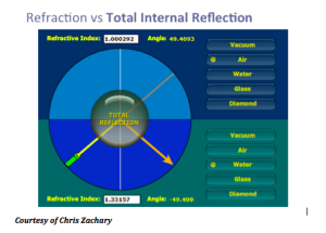
Another concept that Dr Ross discusses is the TRASER (Total Reflection Amplication of Spontaneous Emission of Radiation). The traser is not a laser, nor an IPL. This is one device with many wavelengths that is tunable, has high peak power and variable pulse duration. This device is actually less expensive than a laser and you can change the dye very quickly. The traser uses total internal reflection. We know that if we take light beyond a critical angle, the light will come back towards the same direction.

Screen Shot 2014-05-26 at 8.00.12 AM

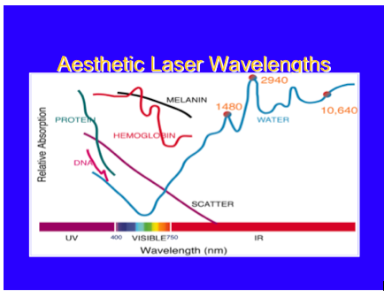
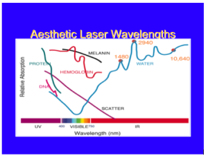
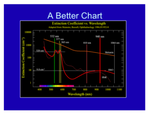
Overall, with lasers in dermatology, you only have to know the chromophore spectra of three targets: blood, melanin, and water. If you know the relative absorption for specific wavelengths for these three targets, you will be a fairly well-armed laser surgeon.

Screen Shot 2014-05-26 at 8.00.00 AM

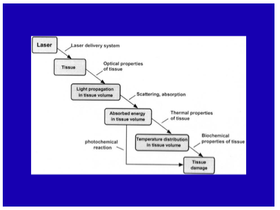
When you model a laser-tissue interaction, you want to look (in your mind) at the way that the light propagates through the skin to the target. You have to get the light to the target. Then, depending on the pulse duration and the wavelength you’re going to have a certain amount of temperature increase leading to a response from that target due to the temperature and time combination.

Screen Shot 2014-05-26 at 7.59.49 AM

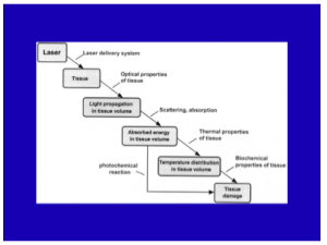
What about skin optics? It’s important that you determine the penetration, the absorption and scattering, and the internal dosimetry.

Laser Tissue Interaction Types

There are different types of interactions. The main type that we use in dermatology is thermal, in that we are basically converting light into heat. Other types of interactions include mechanical, chemical, and plasma. Dr Ross feels that one of best ways to learn about laser tissue interaction is through laser hair reduction. The laser comes down, and a certain amount of the light is going to reach the hair bulb, based on the optical properties of the skin. Typically for 1064nm, about 30 percent of the light is going to get about 3mm down which is the typical depth for a hair follicle. Depending on the pulse width, the wavelength, and the fluence, you will have a certain temperature elevation of that hair bulb and some of that heat is going to diffuse to the surrounding skin. So long as the temperature and the time combination is relatively small, you won’t have too much collateral damage—you will only damage the hair follicle.

Selective photothermolysis was a formal termed coined by Dr Rox Anderson 32 years ago. What it says is that if you have the right wavelength, the right pulse duration,  the right target, and sufficient energy,  you will achieve extreme localized heating. This really revolutionized the way in which we treat vascular and pigmented lesions.

As a dermatologist, you should always remember the graph below, where the wavelength is on the X axis, and you have the relative chromophore absorption in the Y axis. If so, you will be a well-armed laser surgeon. Just like a neurologist looking at an EEG and a cardiologist looking at an EKG, this should be second nature to you.

Screen Shot 2014-05-26 at 7.59.36 AM

How can we exploit laser physics? An example of this is a scar. When we have a scar, there is usually some feature that makes it different from the surrounding skin. You can take a laser and exploit that, whether its redness or pigment in the scar.

This is the temperature equation that basically tells us how hot targets get…the temperature elevation of any target is proportional to the relative absorption of that target for that wavelength of light times the energy div over by a constant. This is very simple concept as its basically energy balanced. Where is the equation??

As you go to longer pulses, the selectivity of the heating becomes poorer but the violent nature of it becomes less.

Fat

Dr Ross states that we are still woefully poor at targeting fat, whether it’s radiofrequency or laser. There are a couple of wavelengths that can be useful for exploiting fat; 1210nm and 1720nm. Those are the wavelengths where fat absorption is in excess of water absorption. However, the ratio of absorption from fat to water is only 1.2 or 1.1 to 1. When we treat a vascular lesion, our absorption rate for the blood is more like 100 to one vs water. So relatively speaking, fat shows poor selectivity, but if you deliver heating and cooling right, there is some selectivity for fat absorption.

Other Key Points

With regards to cooling and heating, we always want to preserve the epidermis and a dynamic cooling device can do so. It is also important to know the photochemistry with regards to chemical reactions and pay attention to these reactions; an example of this can be seen with patients who have previously been on Gold.

Microwaves and Radiofrequency

Microwave and radiofrequency devices are becoming increasingly commonplace within our armamentarium. They rely on heating water, the resistance of water molecules turning causes heat. The  microwave device is around 6GHz. Problems like underarm sweat can be treated with systems like MiraDry. Basically what happens is that the microwaves come down and there is a discontinuity of the dermal-fat junction and it heats up the sweat glands about 4mm below the skin. MiraDry offers another application in clinical practice and can complement other procedures. It also has little downtime.

We are going to begin to see more and more radiofrequency (RF) reactions. It is important to understand SAR, i.e., the specific absorption rate at which energy is absorbed when exposed to an RF electric field. By delivering the right time and the right temperature, one can establish very nice reliable heating of the skin.


MauiDerm News Editor- Judy Seraphine


Pediatric Dermatology: What we’ve learned that has changed the way we think about and practice medicine

Sheila Friedlander, MD and Ilona Frieden, MD

Part 1: Look What the Wind May Have Blown In

Dr Friedlander began this session with a case study….A child presents to your office with an unusual rash.  The pediatrician called it maculopapular,  others migth call it polymorphous. The rash is papular ,  erythematous and fairly generalized.  Dr Friedlander states that most of the time when a child comes into your office with this type of rash, the history is often not that helpful. Be sure to ask about systemic signs, fever, or anyone else sick in the house.  Obtain a drug exposure history. . What is it reasonable to think about? Most of the time, it is a virus. Enterovirus is a reasonable possibility When in doubt, some physicians will blame EBV, especially if the child has had amoxicillin. Occasionally, it could even be measles with this sort of polymorphous eruption,  so it is important to look for cough, coryza, and conjunctivitis. It could also be Parvo virus so check to see if the  child had “slapped cheeks” appearance at any point, or a lace-like appearance to the body rash. . This particular child has had high  fevers for four days and the family is starting to get anxious.  The patient is fussy and his  lips are cracked. The parents also inform you that the child took amoxicillin two days ago and some NSAIDs yesterday. What do you think this is?  Dr Friedlander again emphasizes that it is important to go through his history of drug exposure;  fortunately  in this case, the rash predates any medications.

When you examine the eyes, you note the conjunctiva are red with a rim or halo of white around the cornea,, which is called limbal sparing—In  the diaper area, there is superficial desquamation that is really faint superimposed on confluent erythema. .  What can cause superficial desquamation in this pattern. What about staph scalded skin? The kids who have staph scalded skin do have intertriginous area involvement, but also often have erythema  in the perioral and periorbital areas along with radiating fissures around the mouth. They may also have blisters or bullae.

When you see a patient like this, you need to think about virus, drug, and staph scalded skin. Remember, this child has a polymorphous rash, has lip and tongue involvement, and a very distinctive diaper-area rash that developed  early in his course. The child also has conjunctivitis with limbal sparing and one enlarged lymph node.

So, what should we be  thinking about? Kawasaki Disease…Dr Friedlander reminds us to consider this whenever we see a child who has had prolonged fever  and a rash.

A 2013 article by Bayers S, et al was published in the JAAD, and is an extremely useful review  for dermatologists interested in  Kawasaki Disease. This disease is a small and medium size artery vasculitis.  One of the diagnostic criteria is fever for more than five days; however, if you have a child that fits all of the criteria at day four, you should move ahead with treatment. The earlier you start intervention, the less likely the child is to develop coronary artery disease. The palms and soles are  often red and swollen, and the aforementioned findings of conjunctival injection along with crusted lips and oral mucous membrane erythema are often present.  Cervical l adenopathy is often present but usually asymmetrical—this is helpful to remember.  If you suspect Kawasaki disease, get an expert in the area to help you manage this patient.

What are other KD  skin findings that are of interest to dermatologists?

  • Micropustular follicular rash (uncommon)
  • Nail discoloration, onychomadesis
  • BCG site rash
  • New onset psoriasis

Kawasaki Disease: Epidemiology

The typical age at presentation is six months to five years of age and the highest incidence of the disease is in Asian countries. In the United States, the highest incidence is among those of Asian ethnicity. If a child has this disease, the risk for a sibling to develop the disease is six to 30 times higher than normal and  for any progeny of KD patients, , the risk for his/her child is two times higher. The recurrence rate is two to four percent.

Why is this disease so important? Coronary artery aneurysms and cardiac disease!

It is extremely important to make the diagnosis because cardiac complications can occur. These include:

  • Coronary ectasia and aneurysms
  • Decreased coronary arterial compliance
  • Myopericarditis
  • Arrhythmias
  • Ischemic heart disease
  • Pericardial effusion
  • Valvular regurgitation
  • Myocardial infarction
  • Sudden cardiac death

Cardiac complications affect 15 to 25 percent of untreated patients.

Treatment for Kawasaki Disease

Again, refer to a specialist to help manage these patients. Treatment can decrease coronary risk by at least a factor of five. Treatment includes IVIG 2gram/kilogram over 12 hours, ASA High (80-100mg) divided qid, then low-dose (3-5mg) around day 14. Corticosteroids may be useful in refractory patients. Biologics, such as infliximab and etanercept, may also be useful. IVIG should be repeated if there is no defervescence at 36 hours.

What causes this disease?

We know that cases tend to occur close to each other temporally and there is a prediliction for winter and spring. It occurs mostly in children and is self-limiting. It takes a  course similar to that of  infectious processes  where  immune-mediated response is apparent. Lots of organisms  have been implicated; yet the etiology remains elusive..

Over the last  few years, researchers have found that a correlation exists between wind currents that track from Asia to Japan which  traverse the North Pacific and the occurrence of Kawasaki disease. There have been three epidemics in Japan in May, March, and April, which correlate with times of highest intensity wind currents The belief is that there may be a wind-borne environmental trigger. Investigators are now utilizing  planes and are collecting samples of air in the middle of these wind currents. Many scientists believe that there is a wind-borne agent that is causing this disease. Stay tuned, we may have more information next year.

Part 2: What is a Birthmark?

In the broad sense, a birthmark is a developmental anomaly that is present on the skin. As you know, they do not necessarily need to be present on the day of birth and they can range from common to rare.

Dr Frieden discusses the six questions that you should ask when you see a child with a birthmark.

  • What is the diagnosis? Is this a port wine stain or is this a premonitory mark of an infantile hemangioma. It is extremely important to pin down the diagnosis.
  • Is there a risk of extracutaneous associations?
  • What is the prognosis/natural history?
  • What (if anything) needs to be done? Are there good treatments?
  • Is there a “window of opportunity?”—an example of this would be infantile hemangioma whereby early intervention can really make a difference if treatment is needed
  • What is the cause?

Dr Frieden has been a pediatric dermatologist for a little over thirty years and she states that parents always ask: “what caused this?”  Previously, the answer was “we don’t know”.  Often times, especially for mothers, they ask themselves if they did something wrong during the pregnancy.

So to answer the sixth question, according to Dr Frieden, “2013 has really been a watershed year.”  Researchers are now using deep sequencing of birthmark tissue, not germ-line DNA in order to understand the genes that cause birthmarks. Most of the birthmarks that appear are post-zygotic somatic mutations. Knowing the cause of the birthmark may help to pave the way for newer therapies.

In 2013 Mutations In….

  • GNAQ cause Sturge-Weber
  • KRAS and HRAS causes of nevus sebaceous
  • NRAS cause of giant nevi
  • GTPase BMS1 cause of familial aplasia cutis congenital (germ-line)

What’s exciting about this? New therapies will be developed to address activating mutations because these are also mutations that can cause cancer. We know that researchers are looking for ways to stop activating mutations in these pathways. So you may ask “how does this affect a birthmark?” Dr Frieden provides an example where it may be very important and that is in Sturge-Weber. In Sturge-Weber, children are generally born with Sturge-Weber and they are neurologically normal, there are exceptions to this; however, Sturge-Weber is really a progressive disease. Children will develop heavy paralysis and seizure disorder despite the fact that they are normal at birth. It may be that if we treated these patients with something that inhibited an activated mutation GNAQ only for a couple of years, for example, we may be able to avoid some of the developmental changes that occur. Scientists don’t really know whether or not this is true, but it is an example of where we may be able to halt progression. This is an incredibly exciting time because, as physicians, we may be able to look parents in the eye and provide them with some information about the cause of the birthmark.

Part 3: Drug Reactions in Kids

This section of the presentation focused on drug reactions.,foc. Why is Dr Friedlander cautious when prescribing  minocycline? There is more and more evidence that minocycline is one of the offenders that can cause Drug Reaction Eosinophila and Hypersensitivity Syndrome (DRESS). It’s important to be aware of this disorder and how frequently we can see it.  It  can occur in one in 1,000 to 10,000 drug exposures. DRESS can be distinguished from other drug reactions in that often times the onset is later, approximately two to six weeks after exposure. It presents with a morbilliform rash, significant facial and periorbital edema, and exfoliative changes. Why is it of concernt? DRESS can affect the liver and other organs and has a mortality rate of up to ten percent. This is a good discussion to follow in that of  Kawasaki Disease because they share a similar differential diagnosis. Patients with DRESS have bilateral, significant cervical adenopathy, there may also be significant facial edema, and  often have what looks like an extensive erythematous, sometimes edematous,  confluent eruption. Husain Z, et al published a very useful review in the JAAD in 2013 regarding clinical perspectives and management and therapeutics for DRESS syndrome.

What are the drugs that we need to worry about?

When considering  DRESS, you want to check to see if the patient has eosinophilia and/or liver involvement because such findings raise the likelihood of life-threatening complications. . The common drugs associated with this problem are outlined below:

Ped 1


Pathogenesis

We now know that there are some genetic polymorphisms for drug metabolism that DRESS patients may exhibit. For many of these patients, there may be a genetic difference in how these drugs are metabolized which may be a cause of the disease.  There are two enzymes that have been implicated, epoxide hydroxylase and glutathione transferase. Another fascinating discovery is the association between DRESS and  human herpes virus (HHV-6), CMV, or EBV. Researchers are really not sure whether or not this a secondary involvement, but some scientists feel that re-activation may play an important role. In fact, there are some studies that have demonstrated that those who receive amoxicillin are worse prognostically because amoxicillin, at least in vitro, can help encourage activation and replication of HHV-6. This is an interesting combination of environmental and immune reactions. A number of specific genetic markers have been identified: these include  HLA-A 3101 in association with  carbamazapine; HLAB-5801 with  allopurinol; and HLA DR3 and HLADQ2 with  carbamazapine that put certain patients at risk.

What organ is likely to be involved if your patient gets DRESS?

Ped 2


Don’t forget to think about the differential diagnosis. Remember that the interval before onset is longer than Stevens Johnson Syndrome and TEN. Organ involvement is also more common in DRESS. These patients also tend to have more facial swelling and symmetrical nodes as opposed to Kawasaki Disease. If you were to perform a biopsy in these patients, you would see a more predominant  lymphocytic infiltration whereas in  Stevens Johnson Syndrome or TEN you would see a predominance of necrosis.

What can you do if you think your patient has had a reaction to DRESS?

First and foremost, you want to discontinue the drug. . You can also do skin or serum testing; this  provides a positive predictive value; however, it is not widely used. Therapy for DRESS includes supportive care, systemic l corticosteroids if there is liver involvement, and avoiding the use of antibiotics or NSAIDs.

Prednisone 1mg/kg/day  is often utilized, and tapered slowly over three to six months. If there is no response, patients can be given IV methylprednisolone 30mg/kg IV qd for three days. Do not stop these kids abruptly as there  relapse can occur. It is also very important to check thyroid function.

Dysphagia

Some experts believe that ysphagia is an early manifestation of DRESS syndrome and it may come before the rash. Why is this important? Many times children will present to  the pediatrician with a sore throat and will be given amoxicillin empirically. With the advent of rapid Strep tests, Dr Friedlander would encourage all pediatricians to utilize these tests before simply prescribing amoxicillin, as we know that there is a suggestion that it may be detrimental to patients with evolving DRESS.. Dr Friedlander also recommends antipyretics with Tylenol versus NSAIDs. Remember to ask if the patient is feeling uncomfortable or having trouble swallowing.

MauiDerm News Editor-Judy Seraphine

Pediatric Dermatology: What we’ve learned that has changed the way we think about and practice medicine (continued…)

Sheila Friedlander, MD and Ilona Frieden, MD

Part 4: Hemangiomas and the Use of Beta-Blockers

Propranolol

In this section, Dr Frieden discusses the use of propranolol for the treatment of infantile hemangiomas. A consensus report, published in the Journal of Pediatrics in January of 2013, discussed the initiation and use of propranolol for infantile hemangiomas. Dr Frieden was a co-author on this publication and believes that it may have an affect on the way in which we practice medicine.  The background to this report is that there was a tremendous amount of diversity among the users of this medication. When there is not a lot of data available, the idea of a consensus is to try to come to a central point with general recommendations.

Is hospitalization needed?

The consensus group, comprised by a variety of specialists, recommended inpatient hospitalization if the patient is less than eight weeks old or less than eight weeks adjusted gestational age, and if the patient has other “high-risk” medical conditions. Patients older than eight weeks should receive outpatient monitoring, using heart rate and blood pressure, with dose escalation. Monitoring should be performed one to two hours after dose escalation as great as 0.5mg/kg/day. In Dr Frieden’s practice, she finds that blood pressure monitoring can be difficult with infants; however, heart rate monitoring is easily doable and just about anyone can do this, including parents.

Is EKG needed?

A consensus was not achieved on the use of ECG for everyone.  One may; however, consider ECG in the following circumstances:

  • HR is below normal for age
    • Newborns (<1 month old), <70 beats per minute,
    • Infants (1–12 months old), <80 beats per minute, and
    • Children (>12 months old): <70 beats per minute.
  • Family history of congenital heart conditions or arrhythmias (eg, heart block, long QT syndrome, sudden death); maternal history of connective tissue disease
  • History of an arrhythmia or an arrhythmia is auscultated during examination

The consensus group developed an algorithm that you can utilize or recommend to your pediatric colleagues regarding the initiation of treatment.

Key Take Home Points

In March of 2014, propranolol was approved for the treatment of infantile hemangiomas at a dosage of 3mg/kg/day. A randomized trial demonstrated a 60 percent clearance; however, in systematic reviews, the response rate of propranolol is in the range of 90 to 97 percent, although not everyone achieved clearance. Significant uncertainty and divergence of opinion still exist regarding the safety, monitoring, and dose escalation of propranolol. If the child is at risk for PHACE, at the minimum, you should perform an echocardiogram before considering initiating propranolol. Remember that the peak effect of the medication, in terms of cardiovascular effects, occur one to three hours after administration. The dose response is most pronounced after the first dose and it is extremely important to recheck the heart rate with dose escalation greater than 0.5 percent mg/kg/day. Hypoglycemia is the most common, serious complication. You should discontinue the medication during intercurrent illness, especially with decreased oral intake.

Topical Beta-Blockers

If you are concerned about hypoglycemia and other potential side effects, topical therapy may be a viable option.

What’s the evidence with regards to timolol? There are many citations supporting the safety of  timolol with minimal to no toxicity reported. Most dermatologists have found it useful for thin facial and hand lesions; however, there are some data that support its use in focal, deep facial lesions. A randomized controlled trial demonstrated that timolol 0.5 percent gel twice a day  was a safe and effective option for small superficial infantile hemangiomas that have not ulcerated and are not on mucosal surfaces.

There have been some recent concerns regarding potential toxicity of  timolol when utilized on ulcerated lesions, mucous membranes, or when used extensively in small premature infants.  as it may be absorbed and lead to  systemic levels of drug that could be problematic. Dr Friedlander and her colleagues are currently conducting a study at UCSD investigating  this issue.

Many experts suggest utilizing the drug sparingly, not more than  one to two drops twice a day, particularly in  micropreemies.

Part 5: Vascular Birthmarks and Overgrowth

A 2013 article by Lee MS, et al published in the JAAD reconfirmed Dr Frieden’s approach to the management of vascular birthmarks and overgrowth. Vascular stain and overgrowth were previously lumped together as “Klippel-Trenaunay.” As far back as 2004, Dr Frieden and her colleagues were able to report that the geographic stains had much higher morbidity and a much poorer prognosis. Many cases that were previously diagnosed as CMTC actually reticulate port wine stain. So, how do we give accurate prognostic information to patients and families?

It is important, as clinicians, to distinguish geographic from blotchy/reticulate stains. Conduct serial leg measurements if stains involve the lower extremity and measure head circumference. It is also imperative to look for dysmorphic features, e.g, syndactyly and facila dysmorphism. If dysmorphic features are present, you should consider rare Vascular Stain/Overgrowth syndromes.

Pediatric Dermatology Summary

  • Don’t forget about Kawasaki Disease—maybe the wind blew it in??
  • Birthmarks aren’t necessarily present at birth- and we now sometimes know the associated mutation
  • Dysphagia is an early symptom in DRESS
  • Propranolol is the drug of choice for most problematic infantile hemangiomas and treatment guidelines exist
  • Timolol for children is A-OK


Psoriasis: Update on Emerging Systemic Therapies

Craig Leonardi, MD

At MauiDerm 2014, Dr  Craig Leonardi, Clinical Professor of Dermatology at Saint Louis University, presented us with the latest data on emerging systemic therapies for the treatment of psoriasis. According to Dr Leonardi, “the psoriasis space is going to move very quickly over the next year/year and a half…”

Dr Leonardi comments that many of our 2nd generation biologics were developed with these cytokines in mind (see below).

Screen Shot 2014-04-28 at 8.08.27 AM


Remember that drugs come and go and many fall by the wayside; what’s important to us, as practitioners, is finding the best medicine for our patients.

Certolizumab Pegol (CZP)

CZP is a peglyated Fab fragment. It was initially approved for Crohn’s disease and subsequently approved for the treatment of rheumatoid arthritis, psoriatic arthritis and ankylosing spondylitis. In 2005, there was a large phase II psoriasis trial of 176 patients with 10 percent BSA and PASI 12. It was conducted in Germany and France to look at CZP for the treatment of psoriasis. There were three treatment arms: 200mg sc qeow, 400mg sc qmonth, and placebo. PASI 75 results demonstrated 75 percent, 83 percent and 7 percent, respectively. PGA results (clear-almost clear) showed 53 percent, 72 percent and 2 percent, respectively among all treatment arms. There were no unexpected safety issues. This data tell us that CZP is both efficacious and safe for the treatment of moderate to severe plaque psoriasis. The fact that it is approved for PsA is also important to patients with PsO/PsA. This data was presented in 2007 at the American Academy of Dermatology and published as a paper in 2012. It appears that currently, UCB, the maker of CZP, has again taken an interest in this medication, as there is some development effort and we should be seeing a psoriasis trial quite soon.

Targeting the IL-12/23 Pathway

Both ustekinumab (IL-12) and briakinumab (IL-23) block the shared p40 subunit. When you block IL-12, you down-regulate a set a cytokines from the Th1 pathway, including INFy, IL-2 and TNF-alpha.  When you block IL-23, you down-regulate IL-17 alpha, IL-17f, IL-6, TNF-alpha, IL-21 and IL-22.

Function of Th17 Effector Cytokines

IL-17a

•Expressed by memory NK and T cells

•Increased in psoriatic skin

•Subcutaneous injection à neutrophilia

•Enhances inflammation

•Enhances angiogenesis

IL-22

•Expressed in high levels by Th17 cells

•Increased in psoriasis (skin and plasma)

•Levels correspond to disease activity

•Induces keratinocyte hyperproliferation (in vivo, in vitro)

•Stimulates keratinocytes to secrete antimicrobial peptides

Ustekinumab is a high-performing drug for psoriasis patients. The data at week 28 in both the PHOENIX 1 and PHOENIX 2 studies, show about 70-79% of patients are achieving a PASI 75; this is a huge achievement for these patients.

Screen Shot 2014-04-28 at 8.08.38 AM

New Development Efforts

Ustekinumab was recently approved for the treatment of PsA, based upon its efficacy data with regards to an ACR 20 response at week 24.

Screen Shot 2014-04-28 at 8.08.46 AM

However, when you compare these numbers with the other TNF antagonists, ustekinumab does not appear to work as well in PsA. It remains to see how our rheumatology colleagues will regard this drug over time. It may be used as a second- or third-line drug for PsA.

Screen Shot 2014-04-28 at 8.08.52 AM


3rd Generation Biologics

There is a down-stream effect from anti-IL-23 blockade feeding into IL-17 and down-regulating IL-22. Many pharmaceutical companies have been studying this pathway; there are two, possibly three, IL-23 inhibitors currently in trial, there are two anti-IL-17 inhibitors, an IL-17 receptor antagonist, and there was an IL-22 blocker that came and went…this is a very rich developmental pathway with many promises.

IL-17 Antagonists

The phase II studies of secukinumab (Novartis) a human IgG1 monoclonal IL-17A antibody demonstrated that the 150mg and 300mg doses were statistically significant as compared to placebo at week 12. This is an incredibly high-performance drug, i.e., in the 300mg dose, between 80 and 90 percent of these patients were achieving a PASI 75. According to Dr Leonardi, “this is absolutely remarkable when you think about the world of chronic inflammation”.

The results of secukinumab’s pivotal phase III data was released this fall at EADV.  Dr. Leonardi reported that the ERASURE study evaluated secukinumab doses of 150 mg and 300 mg in a placebo controlled study.  PASI-75 response rates in the secukinumab groups reached a peak at week 16 (80% – 90% range).  IGA scores of “clear or almost clear” were reported in the 60% to 75% of patients. Treatment benefit was maintained through week 52 on q4 week injections following an induction period. Other phase 3 trials included the FIXTURE study included etanercept as an active control.  In this comparator study patients showed a more rapid response to secukinumab vs etanercept (3 vs 8 wks) to achieve a PASI 50 and greater number of patients achieving PASI 90 (72.4% vs 41.5%). The third secukinumab Phase 3 trial SCULPTURE compared fixed-interval q4 weeks maintenance dosing with an “retreatment-as-needed” regimen. Data from follow-up to week 52 showed fixed interval therapy with SEC 300 mg and 150 mg q4wks sustained significantly greater PASI 75, 90 and 100 clearance rates over 1 year compared to “retreatment-as-needed”. “Retreatment as needed” is not a good regimen for any biologic”, says Dr. Leonardi.

We haven’t seen any of the safety results from the secukinumab phase III trials; however, the drug was well tolerated overall according to the phase II data according to Dr. Leonardi.

Ixekizumab (Lilly), is another anti-IL 17 monoclonal antibody, is another high-performance skin-clearing drug based upon its phase II data. Studies looking at 10, 25, 75 and 150 mg demonstrated that at the two highest doses, 80 percent of patients were at the PASI 75 range. There were no serious adverse events in this trial and the drug had a “remarkable safety profile” according to Dr. Leonardi.

The biostatisticians at Lilly, the makers of ixekizumab, utilized Youden’s Index to create a sensitivity and specificity assay. They determined that if they looked at the PASI 50 response at week four, they could make accurate predictions on the chances of success downstream. The likelihood of achieving PASI 75 and PASI 90 correlated with the PASI 50 response.

Brodalumab (Amgen) is an anti-IL-17 receptor antibody, which blocks not only Il-17A but also IL-17E and IL-17C.  It is another high-performance drug for the treatment of psoriasis. The phase II studies, which were completed some time ago, demonstrated positive efficacy results along with a good safety profile. The long-term maintenance of clinical response with brodalumab has been demonstrated through week 96 of an open-label extension study. The one caveat with this open-label extension study is that it is an observed analysis, not an intent-to-treat approach to the data. Remember that the patients who came out of the trial are those who are not doing well so as you move through the trial, the population will continue to improve.

Screen Shot 2014-04-28 at 8.09.04 AM

Positive results were also seen with sPGA over 96 weeks.

Interleukin 23

Remember that the p19 subunit is NOT shared; therefore, an anti-P19 molecule will block just IL-23. Tildrakizumab, a novel anti-IL23p19 monoclonal antibody, demonstrated good results in a 16-week phase IIB trial. There were four doses at weeks 0 and 4 (5mg, 25mg, 100mg, and 200mg) versus placebo.  PASI 75 was achieved in 35, 65.5, 67.1, and 76.2 percent of patients, respectively versus 4.9 percent in the placebo arm and PASI 90 was achieved in 11.9, 24.4, 38.2, and 51.2 percent of patients versus 2.2 percent in the placebo arm. Tildrakizumab appears to be generally safe and well tolerated.

Small Molecules

Apremilast (Celgene)is a novel, small molecule that inhibits PDE-4. It has a variety of immunosuppressive effects, in that it reduces TNF-alpha, IL-2, IFN-γ and several leukotrienes. In Phase III studies (ESTEEM I and II), apremilast (30 mg BID) has achieved a PASI 75 rate of 33 percent. Importantly, scalp, nail and pruritus scores were superior in the apremilast patients compared to controls. Phase III results also demonstrated greater improvements from baseline Dermatology Quality of Life Index versus placebo.  The majority of adverse events (AEs) were not serious and included mild gastrointestinal side effects: nausea 16% and diarrhea 19%.

According to Dr. Leonardi, apremilast also has phase III PsA results that are statistically significant versus placebo although not quite as robust as the TNF inhibitors.  (FDA approval for apremilast for the treatment of PsA was announced during the winter AAD).

Tofacitinib (Pfizer), a novel, oral JAK inhibitor approved for the treatment of rheumatoid arthritis, is currently being investigated as a treatment for psoriasis among other indications. At week 12, PASI 75 response rates were significantly higher for all tofacitinib twice-daily groups: 25% (2mg), 40.8% (5 mg) and 66.7% (15 mg), compared with placebo (2%). It appears to work about as good as methotrexate. Of note, in rheumatoid arthritis, the FDA approved only the 5mg dose. The phase II safety data reported five serious adverse events (SAEs), three of which (atrial fibrillation, pyelnephritis, and urosepsis) occurred in one patient on the 2mg BID dose. There was a mild increase in Hgb levels that was greater in the 15mg BID group. There was a transient decrease in PMNs in the 5mg and 15mg BID groups and there were dose-related increases in both HDL and LDL. In the rheumatoid arthritis trials, the FDA found that the numerical increase in malignancy incidence over time was of particular concern and is consistent with a scenario where increasing exposure to tofacitinib increases the risk of malignancies. The FDA also stated that opportunistic infections, including TB, were “not uncommon.” In the RA program, 14 out of 15 patients who died due to infection were on tofacitinib. There were 34 patients with opportunistic infections, all of whom were on tofacitinib. Despite the immunological differences between rheumatoid arthritis and psoriasis patients, this is remarkable data and something that we, as dermatologists, need to pay particular attention to.

Conclusions

In conclusion, we can see an explosion of new biologics in the marketplace with most of the drugs targeting the IL-23 pathway. We still have an unresolved issue with regards to major adverse cardiovascular events (MACE).  Some feel that there is a signal at the IL-12/23 blockade level, it will be interesting to see if the MACE signal populates the IL-17 blockers…there may be more information coming out on that.  We are also seeing a wide variety of novel small molecules demonstrating moderate to robust efficacies and the safety profiles for some of these products are promising.  There has also been a move to explore the JAK inhibitor in a topical formulation to treat psoriasis.

Psoriasis patients are now the “model” of choice for chronic inflammatory disease and we are getting these drugs earlier now than ever before.  Remember that many of the drugs have fallen by the wayside for efficacy and/or safety issues. We need to select drugs that are good for our specialty and for our patients. As dermatologists, we need to consider that short- and long-term safety issues do exist; however, the benefit/risk ratio appears to be favorable overall.

Our treatment paradigm has shifted from a stratified approach, i.e., patients must fail the previous “step” of therapy before initiating a more “aggressive” therapy to an approach whereby the choice of therapy depends upon individual patient characteristics.


MauiDerm News Editor- Judy Seraphine