Pediatric Dermatology: When to Suspect a Genetic Disease and How to Make a Diagnosis

James Treat, MD

In this presentation, Dr James Treat, an expert in pediatric dermatology, discusses genetic diseases in dermatology and provides us with information on how to make an accurate diagnosis.

Dr Treat reminds us that it is impossible to memorize all of the genetic diseases. As dermatologists, we can identify unusual cutaneous findings, i.e., describe the skin, as this can be helpful in making a genetic diagnosis. Remember that pathways can help you understand the disease(s). If you wonder about a genetic diagnosis, always ask yourself these four important questions:

  1. Does your patient have two or more unusual findings?
  2. Is there is a family history of similar findings?
  3. Is the child developing normally?
  4. Does the child have dysmorphic features?

Case Study Example

A child presents to your office with congenital red patches. What are they? What should we do about them?

  1. Does your patient have two or more unusual findings?—Yes, headaches
  2. Is there is a family history of similar findings?—Two uncles and his father have similar findings and he has an uncle with anyeurism
  3. Is the child developing normally?—He is having some difficulty in school
  4. Does the child have dysmorphic features?—No

If you have a patient who has multiple unusual findings OR multiple family members with one unusual finding, use your resources!

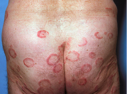
Technology provides us with an abundance of resources. When we have no idea or no specific search terms, we can use Google; however, we must be cautious in its use. In order to weed out non-scientific website answers, use Google Scholar. Does hand, foot, and mouth affect the buttocks? We can use Google images to search; yet, be aware that many images are mislabeled. OMIM can help when your patient has at least two unusual diagnoses and Genetests.org can aid if you’re wondering if a genetic test is available.

Capillary Malformation-Arteriovenous Malformation Syndrome (CM-AVM)

Dr Treat feels that this is something that is probably under-recognized. It’s a RASA-1 mutation that is associated with multiple capillary malformations that are inherited autosomal dominantly. There is usually a family history of multiple people with these red patches.

Clinical Pearls:

  • They look like café-AU-laits, but they are red-purple
  • They may all be nascent AVMS, so be careful using a laser

Remember that some patients have AVMs internally; they can be anywhere in the central nervous system and early screening for AVMs can be lifesaving. Given the association with CNS/spinal AVMs, the potential recommended work-up would be to consider an MRI/MRA.

Case Study Example

A patient presents with congenital red patches. He has a very large head with hydrocephalus. There is no other family history and he is developing normally. Does the child have dysmorphic features? Yes, he has a very large head and his 2nd and 3rd toes are fused (syndactyly). If we use our resources (Google Scholar or Google), we will find the name Macrocephaly-Capillary Malformation (and in small writing macrocephaly-cutis marmorata telangiectatica congenital CMTC).. M-CMTC was the name of this disease for a very long time, and went under-recognized because the skin finding is NOT CMTC it is a more recently described entity called a retiulated port wine stain. As dermatologists, we need to be able to tell the difference between the vascular formations.

Associations:

  • Macrocephaly
  • CM which tends to fade over time and tend to be more evanescent
  • 2nd-3rd toe syndactyly
  • Developmental delays

How do we diagnosis M-CM?

This disease was recently linked to disruption of the PI3k-AKT signaling. Findings indicative of M-CM are macrocephaly plus capillary malformations and two of the following:

  • Asymmetry of overgrowth
  • Developmental delay
  • Midline facial capillary malformations
  • Neonatal hypotonia
  • Syndactyly/polydactyly
  • Frontal bossing
  • Joint hypermobility
  • Hyperelastic skin, and hydrocephalus

This patient was diagnosed with hemangioma with arrested growth. How do we make the clinical diagnosis to differentiate hemangioma from capillary malformations and other vascular malformations? Look for red papules, ulceration (commonly seen in hemangiomas in this area), and large telangiectatic blood vessels as well as a peripheral rim of vasocontriction.

Case Study Example

Your patient presents with exuberant warts on the hands. With widespread, severe warts, we ask ourselves if there could be genetic immunodeficiency. Does your patient have other unusual findings? Yes, he has severe atopic dermatitis and always seems to be sick. There is no family history of similar findings, no dysmorphic features, and the child is developing normally.

Combined Immunodeficiency Associated with DOCK8 Mutations

DOCK8 was recently described as a cause of immunodeficiency that leads to severe viral infections, including the infections that we see as dermatologists, as well as upper respiratory tract infections and severe atopic dermatitis.

Most patients with severe atopic dermatitis and a few warts do not have anything wrong with their immune system; however, if you see a patient with terrible warts and terrible atopic dermatitis, you should think about having that patient see an immunologist.

Case Study Example

You notice congenital tan patches on your patient. What are they? Does your patient have other medical problems? No, and it’s only one tan patch. Is there a family history of similar findings? No. Is the child developing normally? Yes. Does the child have dysmorphic features? Yes, a large head.

This patient has neurofibromatosis Type 1 (NF1-). How do we diagnose this? We need two or more of the following: (Note that several of these findings are dermatologic)

  1. Six or more café-AU-lait spots 1.5cm or larger in post-pubertal individuals, 0.5cm or larger in pre-puberatal individuals
  2. Two or more neurofibromas of any type or one or more plexiform neurofibroma
  3. Freckling in the axilla or groin
  4. Optic glioma (tumor optic pathway)
  5. Two or more Lisch nodules (benign iris hamartomas)
  6. A distinctive bony lesion: dysplasia of the sphenoid bone or dysplasia or thinning of long bone cortex
  7. A first-degree relative with NF-1

What if you are sure that your patient has NF-1 and the testing is negative? You can also test for SPRED1.

SPRED1 is a novel mutation in the same pathway regulated by Neurofibromin, It is part of the SPRED/SPROUTY family which regulates MAP kinase activity (as does Neurofibromin). Most patients reported were adults and did not have other sequalae of NF-1. These patients have some increased risk of cognitive and developmental behavior abnormalities.

What is your workup in a healthy ten year-old with an isolated congenital flat tan patch?

  1. Send blood to evaluate for GNAS1 mutation
  2. Reassure
  3. Send blood to evaluate for NF-1 mutation
  4. Send blood to evaluate for SPRED1 mutation

These consults can be frustrating because we don’t always know what else can be doing on. Segmental pigmentary disorders (block-like, midline cutoff, isolated) have clinical characteristics that are reassuring as there is normally nothing else going on. Other diagnoses to be considered:

  • McCune Albright (typically multiple darker patches along thick lines of blaschko)
  • Neurofibromatosis (typically >6 oval smaller patches)
  • SPRED1 (same clinical café au lait macules as NF1)
  • Speckled Lentiginous nevi (by early childhood can often see the speckling within the larger tan patch)

Conclusions

You, as the dermatologist, have the power of accurate skin description. Look for capillary malformations, warts that are a bit too exuberant and café-AU-lait macules versus other pigmentary disorders. If you suggest a genetic disease, ask the important questions and utilize the resources that technology has provided us.


MauiDerm News Editor-Judy Seraphine

Update in Cutaneous T-cell Lymphoma (CTCL)

Alain H. Rook, MD

At MauiDerm 2014, Dr Rook provided the audience with an update on CTCL, a challenging disease in the field of dermatology. Dr Rook, one of the world’s leading authorities on CTCL, is a professor at the Perelman School of Medicine at the University of Pennsylvania.

There is one scenario that unfortunately occurs all too often with patients who present with erythroderma—you must be absolutely sure of the diagnosis before you administer therapies (anti-TNF agents, ustekinumab, cyclosporine, etc) that will blunt the immune response. This is extremely important because you do not want to administer these agents in patients with CTCL as they can lead to rapid disease progression.

Diagnosis of Erythroderma

In an erythrodermic patient, dermatologists should be aware that a single biopsy is inadequate in up to fifty percent of cases due to the absence of critical diagnostic features.

What do we look for in a biopsy?

Screen Shot 2014-10-26 at 8.23.38 PM

When looking at a biopsy, we look for well-known epidermotropic morphology. We also look for clusters of cells. Another critical finding is the lining up of some large lymphocytes at the dermal/epidermal junction. The malignant T-cells are being recruited towards the epidermis by chemokines, with the “sentinel sign” a characteristic that is more often found in erythroderma than is epidermotropism. We also need to look for large atypical cells in the dermis occasionally in association with eosiniphils. Eosiniphilia, together with erythroderma, should not equal pityriasis rubra pilaris or psoriasis, but is more characteristic of erythroderma associated with either atopic dermatitis or cutaneous T-cell lymphoma (CTCL).

In up to fifty percent of cases, there is a high frequency of non-diagnostic histology in Sezary syndrome, the leukemic variant of CTCL. Published studies have observed chronic dermatitis in 33 percent of biopsies from Sezary syndrome and no evidence of CTCL in 25 percent of biopsies from these patients. “Non-specific cutaneous histopathologic findings are common in Sezary syndrome.” (Trotter MJ. J Cut Pathol. 1997)

In addition, we like to study both the skin and the blood and determine if there is a dominant T-cell clone. In particular, if you see matching clones in the skin and blood, that’s usually typical of CTCL, but not always. Autoimmunity can be associated with dominant clones; however, you typically do not see the identical clone in the skin and blood in a patient with autoimmune disease. This is much more characteristic of CTCL or a T-cell lymphoma.

Flow cytometry is absolutely critical to perform on an erythrodermic patient in whom you would like to rule out CTCL. Prior to about ten years ago, the gold standard was looking for the loss of CD7 on CD4 T-cells that is known to occur in about 50 percent of leukemic patients. Now, we typically look for loss of CD26, occurring in greater than 90 percent of leukemic patients in our experience. This is important because in more than 80 percent of active erythrodermic CTCL cases, you will see active peripheral blood involvement or leukemia. This is a critical test to perform in a highly skilled laboratory. Many of the public laboratories that are not associated with academic programs do not monitor for loss of CD26.

Flow Cytometry

  • More useful than Sezary prep
  • Confirms presence of blood disease
  • CD4+/CD7- or CD4+/CD26-
  • False positive increase in CD4+/CD26- T-cells in atopic dermatitis
  • > 20% circulating lymphocytes that are CD4+/CD26- highly suggestive of Sezary syndrome

Don’t forget about physical examination, i.e., palpate lymph nodes.

It’s important to remember that when you’re treating Sezary syndrome, you should NOT use:

  • Cyclosporine
  • Azathioprine
  • Mycophenylate mofetil
  • Anti-tumor necrosis factor agents
  • Anti-IL-12/23

The reason that we cannot use the therapies mentioned above is because the cellular immune response is absolutely critical for these patients. Combined immune potentiators for advanced CTCL have been shown to produce high response rates. These include combinations of interferons, bexarotene (or an RAR specific retinoid), GM-CSF, and photopheresis. We also attempt to debulk the skin with PUVA or total skin electron beam.

It is noteworthy that a 2011 paper demonstrated that psoralen plus UVA light can be associated with the clearing of peripheral blood disease in advanced CTCL. (Raphael BA, et al. JAAD.2011;65(1):212-214.) In fact, three out of the five patients presented in this series cleared their blood when PUVA was added to their regimen. And we certainly can accomplish the same in the majority of patients who receive full dose total skin electron beam.

Why is the skin involved in T-cell lymphoma?

Malignant T-cells are very high expressors of cutaneous lymphoid antigen (CLA). When the cutaneous endothelium is activated by interferon gamma or tumor necrosis factor, you observe upregulation of E-Selectin. The cells then begin to adhere and roll very slowly. The malignant cells are also very high expressers of CCR4; this is important because CCR4 is the chemokine receptor for CCL17 (TARC) which is manufactured in the epidermis and which is responsible for attracting the cells into the skin.

Screen Shot 2014-10-26 at 8.23.56 PM

Phase I trial data demonstrate that KW-0761, an anti-CCR4 monoclonal antibody with enhanced antibody-dependent cell-mediated cytotoxicity (ADCC) currently in phase III trials for leukemic forms of CTCL, is both efficacious and safe.

Treatment of CTCL

Alemtuzumab, which is particularly useful for leukemic CTCL in the absence of bulky nodal disease, targets CD52 on T-cells, B-cells, and monocytes, which at high dose, causes patients to become severly immunodepressed. In 2007, Maria Bernengo published a clinical protocol for the use of low-dose alemtuzumab.

  • 3 mg days 1 and 3
  • 10 mg days 5 and 7
  • Continue 10 mg every other day until Sezary count < 1000/ mm3
  • Monitor Sezary count Q2 weeks for one month then Q month
  • If Sezary count exceeds 2000/ mm3 resume treatment
  • High response rates of non-transformed disease
  • Low infection rates

Alemtuzumab is extremely effective at this dose and Dr Rook has never seen a serious or life-threatening infection at his institution. But, nevertheless, patients still require prophylaxis against PCP, fungal infection, and against reactivation of Herpes and varicella zoster.

Histone deacetylase inhibitors are very effective for advanced disease, particularly romidepsin. Romidepsin is a parenteral agent that produces responses in advanced disease in about fifty percent for both tumor stage disease and refractory Sezary syndrome. It is generally pretty well tolerated unless the patient has serious cardiovascular disease. While vorinostat is easier to administer because it is oral, patients don’t tend to like it because of unpleasant side effects including diarrhea and fatigue.

Brentuximab vedotin is an agent that is likely to be useful for large-cell transformation and is under investigation. It is currently FDA-approved for CD30-positive lymphomas, ALCL, and relapsed Hodgkin’s disease. This drug is a fusion protein that contains an antibody directed at CD30. This is important because CD30 gets upregulated in large-cell transformation. Appended to it is monomethyl auristatin, a tubulin toxin. In view of its affinity for CD30, brentuximab can be administered to patients with refractory large-cell transformation. Adverse effects can include peripheral neuropathy.

Early Disease

Screen Shot 2014-10-26 at 8.24.04 PM

This is the typical arciform distribution of early disease. The fact that we can see clearing in the center is highly suggestive that the host immune response is playing a role in keeping this somewhat under control. The survival rate of patients with stage 1a disease when treated is similar to that of the general population. It is important to note that patients who present with plaques have a worse prognosis than patients who present with patches, these patients need to be treated vigorously and immediately.

Treatment of Stage T1 (patches or plaques on less than 10 percent of the skin surface area)

  • Potent Topical Steroids
  • Nitrogen Mustard (0.01-0.04% ointment)
  • Carmustine (0.03-0.04% ointment)
  • Narrowband UVB
  • PUVA
  • Topical Retinoids (Bexarotene, Tazarotene)
  • Imiquimod

A novel mechlorethamine 0.016% gel (Valchlor) was FDA-approved in 2013 for the treatment of CTCL. Dr Rook advises that this compound be used with caution because it can be irritating. When using a topical chemotherapeutic, Dr Rook prefers to use it nightly in an effort to avoid drug resistance. However, because Valchlor is irritating, the recommendations are to use it every other night and perhaps in conjunction with topical steroids to prevent/reduce irritation.

Interferon Alpha and Antitumor Effects

Interferon activates anti-tumor cytolytic cells and inhibits the growth of malignant T-cells. It also inhibits the production of Th2 cytokines. We have also found that response rates are greater when used in combination with PUVA, retinoids, photopheresis, and/or interferon gamma. In order to minimize the side effects of interferon, Dr Rook utilizes a low-dose interferon. Nevertheless, there is a dose related effect with higher doses being more efficacious but are less well tolerated.


Treatment Protocol

  • Initial Dose             5-2.5 million Units SQ        3 x per week
  • Increase to:             5-7.5 million Units SQ           4-5 x per week
  • Typical Dose:             5-5 million Units SQ           every other day        


Bexarotene

Bexarotene induces apoptosis of malignant T-cells. Keep in mind that not all patients will respond; there is only about a forty percent response rate due to the likely reason that the critical RXR receptors may not be expressed at the cell surface of the malignant population in all patients. Dr Rook recommends using bexarotene in combination with interferon, PUVA, and/or photopheresis in an effort to prevent resistance. If you are treating patients using bexarotene, they will require intensive control of hyperlipidemia, i.e, use of a statin and/or other lipid-lowering drugs. Bexartone also produces central hypothyroidism by suppressing TSH so thyroid function should be measured with free T4 (not TSH). Bexarotene may be more difficult to use in diabetics and patients with hyperlipidemia.

Imidazoquinolines

These particular drugs are powerful Toll-like receptor agonists and are therapeutically active for CTCL. Many patients may not respond to imiquimod. Dr Rook prefers to use the Zyclara Pump (3.75% Imiquimod) as it is easier to control the amount dispensed as well as easy to spread.

Resiquimod, a combined TLR 7 and 8 agonist, is at the first stages of the pipeline. It has a bioavailability ten times greater than imiquimod and potency up to 100 times greater than imiquimod. A one-gram application induces a systemic interferon alpha response. Dr Rook is conducting a phase 1 trial with resiquimod and has found that patients get systemic absorption, activation of circulating antigen-presenting cells, and widespread clinical responses. This appears to be a very exciting drug that clears treated lesions and induces the resolution of distant lesions.


MauiDerm News Editor-Judy Seraphine

Melanoma Clinic: Lunch, Lesions and Lessions: Part 2

Part 2

Hensin Tsao, MD, PhD

Keith Flaherty, MD

Ilona Frieden

Philip LeBoit, MD

Case 2

In early August of 2007, a 47 year-old patient presented to her PCP with concerns of an atypical appearing lesion on the left lower leg. She was referred to a dermatologist. On August 14th, the biopsy showed 0.97 mm, Level IV, SSM, four mitoses, and non-ulcerated. A wide excision was performed and there was no residual melanoma and no further surgery was recommended. ‎In April of 2009, the patient noted a small cutaneous mass in scar line on the lower left leg.  In June, the patient underwent wide local excision and sentinel lymph node biopsy. The results showed two out of three positive sentinel nodes, the largest metastasis was 4mm; and (+) extracapsular extension.  On July 29, 2009, she underwent (L) inguinal LND and sartorius muscle transposition flap.  Zero out of seven nodes (including Cloquet’s node) were negative.   There were a total of two out of ten positive nodes. She received adjuvant interferon. Unfortunately, two years later, a PET-CT showed an increase in the number of FDG avid lesions in the leg and inguinal and pelvic adenopathy. She’s now considering systemic treatment.

What specimen should be used for determining the patient’s BRAF status?

  1. Primary melanoma FFPE
  2. Primary melanoma FFPE and blood DNA for comparison
  3. Sentinel lymph node metastasis
  4. Soft tissue metastasis from leg

The general guidelines are to take the most recent evidence of advanced disease. Concordance is very important. Do you know that the lesion in the lung is identical to the lesion that presented five years ago according to its genotype?

SNaPshot panel reveals BRAF WT, NRAS and Q61R mutation. On March 21, 2011, she received high-dose IL-2 therapy. In June of 2011, the PET/CT revealed increased FDG uptake in pre-existing left leg subcutaneous nodules and an increase in size as well as FDG uptake in bilateral inguinal lymph nodes. In August, she enrolled in a clinical trial of bevacizumab and ipilimumab. A CT C/A/P performed in April of 2012 revealed an increase in the size of the largest lung mestastasis (15mm to 20mm) and a new 1.6cm liver metastasis. MRI of the brain was negative. June 8, 2012 was her first day on 10-017, a phase 1 study of a CDK4/6 dual inhibitor. By the end of the July, CT C/A/P revealed disease progression in the lung and a new, solitary brain metastatsis measuring 4mm; the disease burden was otherwise relatively stable. In August, she underwent stereotactic radiation to the solitary, 4mm brain mestastasis. October 23, 2012 was cycle one/day one of BKM120/MEK162 trial # 11-308. She remained stable for four months.

Screen Shot 2014-10-26 at 8.18.34 PM

One of our greatest challenges is that NRAS is not something that we can target with a drug. Indirect strategies; however, may work.

Screen Shot 2014-10-26 at 8.18.48 PM

MEK inhibitors in NRAS-mutant melanoma have some efficacy. It’s not comparable to what a MEK inhibitor or BRAF inhibitor can do in BRAF-mutant cancer, but it is certainly a suggestion that may perturb these tumors.

Acne: Clinical Pearls

James Treat, MD

Dr James Treat provides us with an outline on the management of pediatric acne…

Acne Type and Age of Onset

  • Neonatal:  Birth to ≤6 wk
  • Infantile:  6 wk to ≤1 y
  • Mid-childhood:  1 y to <7 y
  • Preadolescent:  ≥7 to ≤12 y or menarche in girls
  • Adolescent:  ≥12 to ≤19 y or after menarche in girls

Neonatal Acne:

  • Benign, self-limited, asymptomatic eruption of erythematous papules and pustules; no comedones
    • Typically localized to cheeks, forehead, and scalp
      • But: can extend into scalp and onto the shoulders (Neonatal Cephalic Pustulosis)

Infantile Acne:

Clinical: Typically starts at around 3-6 months of age up (as young as 6 weeks) and usually lasts 6-12 months

  • Clinically this looks like adolescent acne:
    • Comedones, inflammatory papules and nodules
  • Scarring potential
  • Harbinger for later acne
  • Most Infantile acne is NOT associated with an endocrinopathy

BUT if on exam you see:

  • Excess Growth
  • Advancement of Tanner Stage
  • Cliteromegaly or enlarged testciles.

THEN: Hormonal workup, consider endocrine evaluation

  • FSH, LH, DHEAS, Free and Total Testosterone, 17 OH Progesteterone , androstendione, and Bone Age

Mid-Childhood Acne

Starts between 1 and 7 years of age.

Clinically this looks like adolescent acne:

  • Comedones, inflammatory papules and nodules
  • Scarring potential
  • Harbinger for later acne
  • This is not an age when there is normal androgen production so acne at this age requires a workup.
  • Clinical exam:
    • Excess Growth
    • Advancement of Tanner Stage
    • Cliteromegaly or enlarged testciles.

Hormonal workup and endocrine evaluation

  • FSH, LH, DHEAS, Free and Total Testosterone, 17 OH Progesterone, androstenedione and Bone Age

Treatment (off label): same as adolescent acne except NO tetracycline derivatives

  • Topical retinoids and Benzoyl Peroxide products often enough
  • Systemic choices: erythromycin derivatives, isotretinoin

Contact Dermatitis

Matthew Zirwas, MD

Dr Matt Zirwas provides us with his top ten take-home points from his workshop at MauiDerm NP+PA in Boston…


  1. Add 10 allergens to the TRUE Test to improve diagnostic reliability dramatically.
  2. OK to patch test on 10 mg of prednisone and all the antihistamine in the world.
  3. OK to patch test on immunosuppressives as long as they still have some rash.
  4. Final read MUST be between 96 and 120 hours after patches are put on.
  5. When reading patches, what you feel is what matters, not what you see.
  6. Most positive patch test reactions don’t matter and you shouldn’t even tell the patients about them.
  7. Negative patch testing is extremely helpful.
  8. If have relevant patch test results, must avoid information overload – tell them the minimum amount of information necessary.
  9. Telling people what they CAN use is much more important that telling them what they can’t use.
  10. Occupational cases almost never turn out well.

Dermatoses of Pregnancy: Key Points and Clinical Pearls

Ted Rosen, MD

1. There are physiologic cutaneous changes associated with pregnancy. These include: linea nigra, melisma, striae, faster growing and harder nails, faster growing hair (followed by telogen effluvium 3 months or so post-partum).

2. Treatments for the physiologic changes of pregnancy rarely work well.

  • Melasma: Combination of retinoid, hydroquinone, glycolic acid peels
  • Striae: Perhaps Fraxel laser or Needling device

3. The immune system changes during pregnancy

  • TH1 cell-mediated immunity decreases: EGW may worsen
  1. Imiquimod appears safe
  2. Thermotherapy is safe
  3. Cryotherapy is safe
  4. CO2 laser is treatment of choice
  • TH2 humoral immunity increases: Lupus may worsen or appear for first time

4. Specific dermatoses of pregnancy have been consolidated into just a few entities:

5. Intrahepatic cholestasis of pregnancy

  • Risk to mother: None (itches)
  • Risk to fetus: Stillbirth, Premature birth, intra-cranial hemorrhage
  • Therapy: Oral ursodeoxycholic acid 15mg/kg/day in QID divided doses

6. Herpes (Pemphigoid) Gestationis

  • Risk to mother: Autoimmune disorders: vitiligo, Grave’s disease, alopecia areata, IBD
  • Risk to fetus: Small for gestational age, premature delivery, blisters (10%)
  • Therapy: Systemic steroids

7. Pruritic Urticarial Papules & Plaques of Pregnancy (PUPPP)

  • Risk to mother: None (itches severely)
  • Risk to fetus: None
  • Therapy: Topical steroids, UVB (sunlight)

8. Prurigo of Pregnancy (PP)

  • Risk to mother: None
  • Risk to fetus: None
  • Therapy: Topical steroids

9. Impetigo herpetiformis (Now considered pustular psoriasis of pregnancy)

  • Risk to mother: Electrolyte abnormalities, especially calcium homeostasis
  • Risk to fetus: Spontaneous abortion, Stillbirth
  • Therapy: Systemic steroids or cyclosporine

10. Excellent review: Dermatol Ther 26:274-84, 2013

10 Pearls from the Basic Structure of Skin

Whitney A. High, MD, JD, MEng

Dr High provided the MauiDerm NP+PA Fall 2014 audience with a comprehensive presentation on the basic structure of skin. Here’s what you need to know…

1. The skin consists of “three layered cake.”  The epidermis an outer protective outer layer.  The dermis is a middle layer that provides “tensile” strength.  The deeper subcutis is insulating fat.

2. The epidermis gets all its nutrition and sustenance from the dermis.  The dermis contains all the “supportive” structures of the skin, such as blood vessels, nerves, and many “adnexal structures.”

3. The epidermis consists chiefly of keratinocytes (“skin cells”).  These cells are arranged in layers to form a “maturing” protective layer that replaces itself every ~28 days:

  • stratum basal = the germinative layer of the skin that divides to regenerate the epidermis
  • stratum spinosum = names for the intraspinous properties that bind the keratinocytes
  • straum granulosum = the granular layer where keratohyaline granules are produced
  • stratum corneum = the “dead” outer layer the provides the most barrier function

4. The dermis is comprised of three main building blocks: collagen, elastic fibers, and “ground substance.  Collagen is the material that provides the tensile strength to the skin.  Elastic fibers provide skin resiliency.  Ground substance facilitates the diffusion of nutrients and oxygen.

5. The “dermoepidermal junction” is where the epidermis attaches to the dermis.  This is also the location of melanocytes that make protective melanin for the skin.  The DEJ is where most nonmelanoma skin cancer invades the dermis, the place where nearly all melanoma originates, and the place where many bullous and “interface” diseases transpire.

6. When confronted with a skin disease, one must ask themselves, “where do I believe that that pathology is occurring?” For example, the pathologic process might be:

  • epidermal – such as the spongiosis (intraepidermal edema) and weeping of dermatitis, or the yeast/hyphae of tinea versicolor/pityriasis versicolor growing in the stratum corneum
  • dermal – such as the histiocytic/macrophagic infiltrate of granuloma annulare, or the neutrophilic inflammation of small blood vessels in leukocytoclastic vasculitis
  • subcuticular – such as the panniculitis of erythema nodosum

7. Being able to predict where the likely pathology is occurring also facilitates the securing of a “representative” biopsy, which is always the responsibility of the clinician.

8. Adnexal structures may be the site of inflammatory pathologic processes (acne, hidradenitis), may be the site of neoplastic processes (sebaceous carcinoma), or these structures may simply behave in an undesired way (seborrhea, hyperhidrosis).

9. Uncontrolled and unchecked growth of certain components of the skin leads to cancer, such as basal cell carcinoma (from basilar keratinocytes), squamous cell carcinoma (also from keratinocytes) and melanoma (from melanocytes).

10. Skin structure and function changes with age, such as dyspigmentation and facial wrinkling, formation of “solar elastosis” (damaged elastic), and increased water loss to the environment.

Genital Ulcers

Ted Rosen, MD

What causes genital ulcers? Are you treating patients who present with genital ulcers? Dr Rosen provides us with his clinical pearls from MauiDerm NP+PA Fall 2014…

  1. Ulceration is defined as loss of epidermis and then some or all of dermis and subcutaneous tissue leading to an “open sore.”
  2. There are many general reasons for ulcers to occur. They include: atherosclerotic vascular disease, non-atherosclerotic vascular disease, infections, inflammation of unknown etiology, neoplasms, and exogenous causes
  3. It is often necessary to use ancillary aids to diagnose genital ulcers; these may include serologic tests (eg. RPR), cultures, and biopsy with molecular technique analysis.
  4. STDs commonly cause genital ulcers. You are NOT likely to see the minor STDs in the USA (eg. Donovanosis, chancroid or LGV). However you may see syphilis and herpes. STDs can be distinguished by lesion size, number, degree of pain, presence or absence of inguinal adenopathy.
  5. Genital herpes due to HSV-2 continues to shed virus for at least a decade after initial infection. Genital herpes should prompt an investigation for other STDs (10+% will be HIV+)
  6. Amebiasis of the genitalia may result from anal intercourse with an individual with amoebic dysentery. Or, it may ensue from fecal contamination of the genital skin.
  7. Neoplasms which cause genital ulcers are >90% likely to be squamous cell carcinoma
  8. Multi-system disease which may be associated with genital ulcerations include Behcet’s Syndrome and extra-intestinal Crohn’s disease. Pathergy helps confirm the diagnosis of Behcet’s disease; biopsy confirms cutaneous Crohn’s disease. Topical TCI may help the latter.
  9. Lichen planus is typically the cause of painful erosions in women, but not in men. Treatment is with ultrapotent topical steroids until healing (~75% of women will heal totally). Alternate treatments include topical TCIs and oral cyclosporine-A
  10. Zoon balanitis is a superficially eroding disorder of men with sheets of plasma cells on biopsy. (Hence the name: balanitis plasmacellularis). Topical steroids treat this.
  11. Unusual causes of genital ulceration can be diagnosed by PE and historical data. These include ecthyma gangrenosum, pyoderma gangrenosum, and calciphylaxis. Beware of EG in neutropenic patients, PG in those with RA or IBD, and calciphylaxis in those with CRF on dialysis.
  12. Exogenous factors causing genital ulcers: self-mutiliation, drugs, vacuum erection device and genital bite wounds. The latter respond best to amoxicillin-clavulanate in high dose.

Dermatology In Review: Part 2

Matthew Zirwas, MD

At the MauiDerm NP+PA Fall 2014 conference, Dr Zirwas reviewed with us some of the key findings in dermatology from 2014…

  1. Coconut oil may help atopic dermatitis
  2. “Supplementing the diet of well-nourished adults with mineral or vitamin supplements has no clear benefit and might even be harmful
  3. UV exposure may reduce all-cause mortality!
  4. Metformin 500mg TID helped 18/25 hidradenitis patients
  5. Rifampin 10 mg/kg once daily + Moxifloxacin 400 mg daily + Metronidazole 500 mg tid for hidradenitis?
  6. Fexofenadine 240 mg bid + Montelukast 10 mg qd helped about ½ prurigo nodularis patients
  7. Is post-op pain due to allergic reaction to metal in implants? Maybe. But not commonly.
  8. Probable efficacy of gabapentin in the improvement of pruritus and quality of life of patients with nostalgia paresthetica (max dose 900mg TID)

Vasculitis and Vascular Lesions Part 1: Clinical Pearls

Ted Rosen, MD

1. Vasculitis is a clinicopathologic process characterized by inflammation of and damage to blood vessels
2. Heterogeneous group of disorders leading to a broad group of named diseases due to….  Different sizes, types and locations of the affected blood vessels and variable tissue response to injury
3.  Basic principles:

  • All age groups (Some types have preference)
  • Caucasians (But other ethnic groups, too)
  • Genetic predisposition (But not strictly inherited)
  • Chronic, relapsing (But may go into remission)
  • Think vasculitis when faced with an…..unexplained multi system disease or progressive organ dysfunction
  • Team approach: Early consultations

4. Classification: best done by size of blood vessel affected

5. A wide variety of tissues can be affected. Scoring charts exist which are helpful to systematically asses these patients.

6. Constitutional signs and symptoms may be seen: fever, chills, sweats, weight loss, fatigue

7. Involvement of, damage to and symptoms relating to: eyes, upper or lower airway, joints, kidneys, lungs, central/peripheral nervous system,  heart and clotting mechanisms

8. Morphology of skin lesions is suggestive but virtually never diagnostic of histologic type!

9. Absence of skin lesions does not rule out any variant of vasculitis, while the presence of any specific type of skin lesion does not necessarily predict the subtype of vasculitis present!

10. There are many clinical mimics to vasculitis. These include embolic phenomenon, exposure to certain drugs, cryptogenic infections, Vitamin C deficiency, and cardiolipin antibody syndrome

11. When biopsy is done, try to utilize a lesion less than 72 hours old to get the most representative histology.

12. Therapy may include: NSAIDS, Dapsone, Colchicine, Steroids, Immunosuppressives: Methotrexate, Mycophenolate, Azathioprine, Cyclophosphamide and Rituximab