Infectious Diseases Update: Part 1: Interesting ID Literature

Ted Rosen, MD

In this presentation, Dr Rosen provides us with new information on infectious diseases that has recently appeared in the literature.

MRSA

A study in the International Journal of Antimicrobial Agents states that minocycline is often forgotten but preferred to trimethoprim-sulfamethoxazole or doxycycline for the treatment of community-acquired methicillin-resistant Staphylococcus aureus skin (MRSA) and soft-tissue infections. (Cunha B. Int J Antimicrob Agents. 2013;42:497). They point out that there is about a thirty percent discrepancy in discordance between the in vitro antibacterial testing and actual clinical effectiveness when it comes to the tetracycline family, this is less so with minocycline.

When treating MRSA, remember that incision and drainage is the most important intervention, if possible. CA-MRSA can be either modestly or severely virulent depending upon the presence or absence of PVL (Panton-Valentine leukocidin).

Typically we have some choices in treatment. We tend to leave linezolid for the last resort and clindamycin often has inducible resistance so we’re left with doxycycline, TMP-SMX, and minocycline. Cunha and his colleagues point out that second only to TMP-SMX, minocycline is the best in tissue penetrability and is equivalent to linezolid in predictable efficacy. Nationwide, only one to two percent of MRSA are resistant to linezolid and minocycline; however, up to twelve percent are resistant to doxycycline.

Because there is a generic minocycline available, the cost issues are largely negated. There are other issues such as hypersensitivity reactions and rare autoimmune phenomena. The authors feel, from a microbiological standpoint, minocycline is a preferred drug.

Herpes

We now have some new therapies available for the treatment of herpes. Herpotherm utilizes thermotherapy for genital herpes. A prospective study conducted in Russia evaluated 32 women. Twenty-one patients received thermotherapy plus acyclovir and ten received thermotherapy alone. Treatment was initiated with the first objective sign of HSV-2. Within one day, symptoms were gone or almost gone, with or without acyclovir as concomitant therapy demonstrating that thermotherapy is beneficial for genital herpes. Although exposure id of short duration (seconds), patients should be warned to expect some discomfort.

Topical zinc sulfate in-vitro inhibits the growth of HSV. Mahajan and colleagues tried various concentrations of zinc sulfate on 100 clinical and Tzanck verified men with genital HSV over a six-month period. They had a very specific treatment protocol:


  • Q5d x 1 mo
  • Then Q10d x 2 mo
  • Then Q15d x 3 mo

Of note, all of the patients had nine or more recurrences per year, i.e., frequent outbreaks of genital HSV. Here’s what the authors found:

Screen Shot 2014-11-23 at 5.20.07 PM

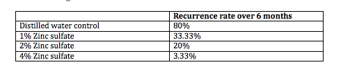
4% Zinc sulfate is a cheap, non-toxic therapy that is easy to use and may be a viable treatment for hard to manage HSV-2. The key is to apply this when there is an outbreak and as it heals, patients should continue to follow the protocol. (Mahajan BB. Indian J Sex Transm Dis. 2013;34:32-34.)

Flat Warts

A study by Li and colleagues, published in the Journal of the European Academy of Dermatology and Venereology evaluated photodynamic therapy with five percent, ten percent, and twenty percent aminolevulinic acid (ALA) in the treatment of generalized recalcitrant facial verruca plana. This was a prospective study of 55 adult patients with bilateral facial flat warts. The two sides of the patient’s faces were randomized to receive ALA 5 percent, 10 percent or 20 percent. There was no placebo control. Patients were irradiated with a 633-nm red light in two 7.5- minute sessions, separated by 30 minutes of non-exposure. This was repeated three times, every two weeks. This study demonstrated that ALA 10 and 20 percent were most efficacious for the treatment of generalized recalcitrant facial verruca plana. In the United States, we have Levulan Kerastick (20% ALA-d). (Li Q, et al. J Eur Acad Dermatol Venereol. 2013 Nov 25. [Epub ahead of print])

Onychomycosis in Psoriatic Patients

A systematic review by Klaassen and colleagues analyzed the literature with regards to the prevalence of onychomycosis in patients with nail psoriasis. They looked at over 700 studies out of which they chose ten based on their criteria. The authors found that an average of 18 percent of psoriasis patients with nail dystrophy have concurrent onychomycosis. There was no shift from dermatophytes to saprophytes or yeast in these studies. Because psoriasis is treated with immunosuppressive agents, this may aggravate any concurrent fungal infection. (Klaassen KMG, et al. J Eur Acad Dermatol Venereol. 2013 Aug 19)

 Take Home Message: It is important that we check for fungal disease in patients with psoriasis nail dystrophy before instituting therapy.

Atypical Mycobacteria/Nontuberculous Mycobacteria

In a case series and epidemiologic study, Falsey and colleagues looked at cutaneous inoculation of nontuberculous mycobacteria during professional tattooing. How does this happen? One to four weeks after the tattoo is placed, it may itch or hurt and there are monomorphous crusted papules or postules that are confined to the tattoo area. At first blanche, we might look at this and think it’s bacterial, but it turns out that it is Mycobacterium either abcessus or chelonae. Clarithromycin, minocycline and ciprofloxacin are all viable treatment options; however, the authors strongly recommend that you send off one the papules for culture and sensitivity because the sensitivities vary widely. Why does this happen? Because the atypical mycobacteria are found as a contaminant in the tattoo ink or sometimes the non-sterile water used to dilute the ink (especially when outsourced from China). (Falsey R, et al. Clin Infect Dis. 2013;57(6):e143-147.)

Take Home Message: Think NTM in new tattoo lesions

Herpes Zoster

Wang and colleagues conducted a population-based retrospective cohort study looking at herpes zoster infection and acute coronary syndrome (ACS), i.e., any event that suddenly decreases blood flow to the heart muscle (heart attack, unstable angina). This study included 59,958 patients with herpes zoster compared to an age/sex- matched cohort of 231,832 non-zoster patients. The authors found that acute coronary syndrome was associated with herpes zoster 25 percent more commonly. This was statistically significant (p=.0001) at both three months and one year after the herpes zoster was resolved. (Wang CC, et al. Br J Dermatol. Dec 6, 2013. e-pub ahead of print)

Take Home Message: Warn zoster patients about the risk of ACS and review symptoms of ACS.

Flood-related Skin Diseases

Flood, one of the most common natural disasters, has contributed to many public health problems. A study conducted in Thailand identified the following flood-related dermatological disorders:

  • Irritant contact dermatitis
  • Webspace tinea pedis
  • Webspace mixed infection
  • Cellulitis (S. pyogenes, Aeromonas)
  • Gas gangrene (clostridium)
  • Fasciitis (Vibrio spp)
  • Traumatic wounds (check cuts for lacerations, punctures, serious digital injuries)
  • Insect bites and stings (mosquito, fire ant, centipede)
  • Animal bites (stray dogs/cats, snakes)
  • Aggravation of pre-existing skin disease due to psychic trauma (psoriasis, atopy/eczema, alopecia areata, urticaria, vitiligo)


Functional Facial Anatomy: A Primer

Sandy Tsao, MD

Where are the safe areas to inject? What can we do in certain areas that we cannot do in others? In order to answer these questions, we need to understand the basic facial anatomy.

Remember that it only takes one complication to understand how significant the facial anatomy can be. As the neuromodulators change and as we become more knowledgeable about treatments, we have a better perspective. When we’re talking about facial anatomy, we’re thinking about the muscles of facial expression that run from the skull to the skin. These muscles are innervated by the facial nerve and are sphincters and dilators of the eyes, nose and mouth. It is key to understand that the wrinkles that we’re seeing are actually perpendicular to the action of the muscle. This is very important for us when we’re thinking about where the lines are and how we would like to get rid of them.

The facial skeleton is composed of fourteen stationary bones and the mandible. These fourteen bones form the basic shape of the face and are responsible for providing attachments for muscles that make the jaw move and control facial expression.

The facial nerve divides into five terminal branches for muscles of facial expression:

  • Temporal
  • Zygomatic
  • Buccal
  • Marginal mandibular
  • Cervical

If you’re ever cutting or injecting into any of these areas, it is critical to think about the insertions and the direction of these nerves.

The skin of the face is supplied by the trigeminal nerve (V), except for the small area over the angle of the mandible and the parotid gland that is supplied by the great auricular nerve (C2 and 3). The trigeminal nerve (V) divides into three major divisions—the ophthalmic (V), maxillary (V2), and mandibular (V3) nerves.

The arterial supply comes from the common carotid artery and it will innervate and branch thoroughly throughout the scalp and the facial structures and drain via the jugular nerve. Why is this significant? Every time we inject, there is always the potential for a hematoma, a bruise or an injury. Understanding where that vasculature is and where the drainage spots are is helpful to minimize side effects.

Muscles

The Temporoparietalis is key to us because of the temporal nerve. This muscle allows us to raise our ears, widen our eyes, and retract our temples. This can be an ideal place to add a filler; however, it is critical to understand that the temporal nerve is a little deeper. Across the face we have the Frontalis muscle that allows us to raise the eyebrows, widen the eyes, and furrow the forehead. We often take advantage of this to minimize and soften the horizontal lines across the face.

In direct opposition are the depressor muscles of the upper face, which include the Corrugator muscles. These muscles interdigitate with the frontalis; they actually displace the brow inferomedially creating the frown. The Corrugator muscles work in conjunction with the Depressor Supercilii that causes medial brow frowning. Last not but not least is the Procerus muscle—a very important muscle because it not only displaces the brow medially, but also creates the horizontal bands that many patients are interested in improving.

Clinical Pearl-When you are influencing one set of muscles, almost always there is another muscle that is acting against it.

The Transverse Nasalis muscle is a muscle is that interdigitates with the opposite muscle as well as the Procerus muscle. This is the muscle that allows for depression of the cartiginous part of the nose as well as drawing the ala toward the septum. This muscle is what we refer to as the “bunny lines.” We want to be very careful with injections here because too low of injections can infect the smile lines. This is one muscle into which we directly inject and try to keep on the higher edge of the muscular complex.

The Orbicularis Oculi is muscle that has two parts—palpebral and orbital. It has two components, each of which need to be thought about because influence of one portion may influence another part of the muscle. This muscle helps us to open and close our eyes, allowing us to form tears. When we’re addressing this muscle, we’re generally dealing with the orbital aspect of the muscle. When injected correctly, this can provide softening of the crow’s feet. If you inject the Orbicularis Oculi too deeply, you can infect the smile by influencing either the Zygomatic Major or Minor or even the Labii Inferioris Superior.

Clinical Pearl-It is pertinent to understand your endpoints because you will have migration of the neuromodulators over a three- to four-month period of time.

Levator Muscles of the Mouth

The Zygomaticus Major helps to raise the angle of the mouth superiorly and posteriorly and helps our mouths smile or laugh. The Zygomaticus Minor runs medially to the Zygomaticus Major and allows the upper lip to displace superiorly and deepens the nasolabial furrow allowing us to make an expression of contempt.

What about the rest of the mouth? The Depressor Labii Inferioris displaces the lower lip inferiorly and slightly laterally. The consequences of which will allow for displacement of the skin downward and lateral pull to the mouth and potentially an uneven smile if the Mentalis muscle is injected to superiorly. The Risorius muscle is one of our elevators and as a consequence it displaces the skin of the cheek posteriorly, it stretches the lower lip, and displaces the corners of the cheek downward and lateral and we see a nice grin.

The Levator Labii Superioris Alaeque Nasi is an important to muscle to know because it dilates the nose and actually raises the upper lip. The Levator Labii Superioris muscle is responsible for raising the upper lip. This is one of the muscles that we like to target when we’re trying to get less of a grin. When a neuromodulator is placed into these muscles, you can see less “gummy” show in the upper lip.

The Orbicularis Oris is also a very critical muscle. It is quite large and has a number of insertions into it. This muscle helps to bring the lips together and protrude forward. The Buccinator muscle merges with the upper and lower lip muscles and helps to compress cheeks against the teeth along with tensing and contracting the cheeks for a nice pucker. Injection into the vertical Rhytids will allow for some softening of the lines created by that muscular action.

The Depressor Anguli Oris helps as a depressor muscle. It depresses the angle of the mouth resulting in an expression of grief. This muscle is often times sought after as a muscle either to relax by a neuromodulator or to uplift by dermal filler. The Mentalis elevates and protrudes the lower lip allowing for an expression of doubt. This is a critical area to avoid surrounding muscles so that the smile itself is not influenced.

The Platysma muscle is a large muscle that helps to shape not only the lower face, but also heavily influences our neck musculature. It results in the depression of the lower jaw, displaces the lower lip inferiorly, and allows for an expression of horror or fright.

Screen Shot 2014-11-23 at 5.14.08 PM

Conclusions

In conclusion, knowledge of the facial muscles is paramount in procedures affecting facial animation. We must understand the relationship of facial muscles and associated nerves and vessels as well as the relationship of muscles and planes throughout the face.

MauiDerm News Editor-Judy Seraphine


Melanoma Clinic: Lunch, Lesions, and Lessons: Part 3

Hensin Tsao, MD, PhD

Ilona Frieden

Philip LeBoit, MD

Keith Flaherty, MD

Case Study

A forty year-old woman presents to her PCP with a lesion that she had removed near her left knee (cryotherapy) about one year ago. She thinks something is returning. “It is a tiny miniscule nodule that appears to be a scar.” This appears to be a dermatological issue and “she wants to see her old dermatologist to have it rechecked.” What is an appropriate next step for the physician?

  1. Refer and document
  2. Refer and follow-up with phone call in 1 m
  3. Refer and call the local dermatologist
  4. Refer and follow up with letters until lesion is removed

If you have a general concern, three certified letters can be a good approach. It is extremely important to follow-through and maintain good communication so that you are not held liable.

Seven months later, the patient returns to the PCP with her parents. She was diagnosed with an invasive melanoma. She didn’t see the dermatologist, whom she was supposed to see last spring, until just recently. The patient was referred to Massachusetts General Hospital.

Melanoma on the left knee showed NMM; 7.5mm thick, ulceration 0.4mm, 12 mits/mm2; no microsatellites. She underwent wide resection and sentinel lymph node biopsy (0/8). One year after the diagnosis, she returns for a follow-up and informs you that she wants to become pregnant. She is now 41 years old and she had a miscarriage several years ago. This scenario is not uncommon and Dr Tsao has had referrals regarding this situation.

What do you tell a 41 year-old woman with a stage IIc (life expectancy 30-50%) melanoma about pregnancy?

  1. Fine to become pregnant
  2. Wait at least 2 years
  3. Wait at least 5 years
  4. Wait at least 10 years

Dr Flaherty comments that the fundamental question that we don’t have a great answer to is whether or not every instance of microscopic metastatic disease would ultimately manifest as overt metastatic disease with time. Or, are there patients who can live chronically, i.e., forever with microscopic disease? We do know of late relapses with melanoma. Dr Flaherty tends to tell patients the suggestion that microscopic metastatic disease will convert to macroscopic metastatic disease and the worst that could happen, in the pregnancy setting, is that you could influence the conversion, in other words, it could happen sooner. It is also important to discuss where patients are on the risk curve. The risk of occurrence falls with time.

The risk of relapse after 10 years is actually not clear because high-risk lesions should have relapsed by then while thin relapses continue to emerge. There are differences in the kinetics of relapse between a thin melanoma and a thick melanoma. The bigger the tumor, the more aggressive it is. If the patient survives to year five or ten, it is less likely that something will happen. In fact, there might be a cross over point, probably around year 10, whereby if you had a thick tumor and you’re still alive, the likelihood of it recurring is quite low.

Pregnancy and Melanoma

There are many case control series of melanoma during pregnancy versus not during pregnancy. A 2008 study found no evidence that pregnancy worsens the prognosis. (Cancer Causes Control. 2008;5:437) Some studies suggest that melanoma during pregnancy may be slightly thicker but survival is not altered that much.

The patient visits with a high-risk obstetrician who clears her to become pregnant. Three months later, she finds out that she is pregnant. How do you follow a pregnant person with a history of melanoma?

  1. Routine post-melanoma surveillance
  2. Routine surveillance with total body photography
  3. Increased surveillance every three months
  4. Increased surveillance with total body photography

Total body photography could be a viable option. There is really no literature on dermoscopy in pregnancy. These patients should be followed very carefully. Regarding post-melanoma surveillance, you don’t need to necessarily change your practice because of pregnancy. There are no great studies that convincingly demonstrate a higher risk of relapse during pregnancy. This is highly anecdotal, yet it is very compelling and memorable when it happens.

At week 17 of her pregnancy, she noticed a small, firm nodule within her melanoma scar. Wedge biopsy showed recurrence of melanoma with neurotropism. Wide resection cleared recurrence and a skin graft was placed.

Management of Melanoma Recurrence During Pregnancy

There are treatment options; however, there are no data for metastatic disease during pregnancy. With regards to immunotherapy, high-dose IL-2, which is a physiologic stress, poses a theoretical risk of abruption (in the setting of hypotension). Ipilimumab is likely the first-line choice. Looking at targeted therapy, BRAF inhibitors showed no risk of teratogenicity in limited preclinical studies and MEK inhibitors are theoretically of greater concern; however, that may not be the case in combination with BRAF inhibitors. Chemotherapy is rarely an attractive first-line option in any case.

Package Inserts

  • Vemurafenib
    • Pregnancy Category D
    • Vemurafenib can cause fetal harm when administered to a pregnant woman based on its mechanism of action
  • Iplimumab
    • Pregnancy Category C
    • Use ipilimumab during pregnancy only if the potential benefit justifies the potential risk to the fetus.

The patient received XRT to the left knee graft area with shielding of the fetus to prevent radiation crossover. At 36 weeks, she underwent a Caesarian section. A histological examination of the placenta did not reveal melanoma. (There have been reports of metastases to the placenta or fetus) One month after delivery she noticed two subcutaneous nodules in the mid-calf distal to the scar site. A biopsy showed metastatic melanoma consistent with in-transit disease. PET/CT showed multiple left lower leg FDG avid nodules, but no visceral metastases. Both radiologically and clinically, she’s beginning to develop at a faster pace.

What would you recommend now?

  1. Isolated limb perfusion
  2. Targeted removal of individual lesions
  3. Genotyping with anti-BRAF agent if BRAF+
  4. Systemic treatment with IL-2 or immune checkpoint antibody

In-Transit Metastases

We lack high-level evidence for the optimal approach in this case. According to the NCCN guidelines, enrollment in a clinical trial is the preferred approach. Options for local therapy include complete surgical excision, intralesional injection (BCG, IFN, IL-2), local ablation therapy, topical imiquimod for superficial dermal lesions, and consideration of palliative XRT for unresectable disease. Isolated limb infusions/perfusions with melphalan could be considered as a regional therapy option as well as the use of systemic therapy. Systemic used to be considered as bottom of the list; however, it is being utilized more and more.

The issue with regional therapy is that it has no impact on disease elsewhere. With regards to intralesional therapy, Weide and colleagues found a high response rate after intratumoral treatment with IL-2 in a phase two study of 51 patients with metastatic melanoma. The 2-year survival rate was 77 percent for patients with stage IIIB/IIIC disease. Efficacy and survival did not differ between patients who had greater than or equal to 20 lesions and patients who had less than 20 lesions. (Weide B, et al. Cancer. 2010;116(17):4139-4146.)

Alexander and colleagues analyzed factors that influenced the outcome in patients with in-transit malignant melanoma undergoing isolated limb perfusion using modern treatment parameters. Between May 1992 and February 2005, 91 patients underwent a 90-minute hyperthermic ILP. There were 62 complete responses (69%) and 23 partial responses (26%) in 90 assessable patients.

The patient underwent isolated limb perfusion with melphalan but unfortunately continued to progress. She continued to progress through multiple experimental treatments over the ensuing year and she expired six years after her initial diagnosis.

Pregnancy and Melanoma-Take Home Points

  • Pregnancy does not appear to compromise survival
    • “Wait time” considerations include:
      • Potential treatment complications during pregnancy
      • Age of patient and desire for children
      • Personal outlook (e.g. religion, “abandoning” children)
      • Availability of oocyte cryopreservation
    • Risk extinction is more obvious for thicker tumors
  • Bottom line (not scientifically based)
    • For patients with multiple DN- watch q3m’s during pregnancy
    • For MIS and thin melanomas- OK to start (need discussion)
    • For melanomas >1mm- wait 2 yrs unless age is major concern
  • Discussion (Push/pull model)
  • Push information-
    • There is the chance of relapse and death (estimate prognosis)
    • Come to an agreement on a course of action
    • There is a tiny chance of transplacental metastasis
    • Pregnancy per se does not appear to influence outcome- gestation cannot be an instrument of guilt or regret
    • Need partnership with high-risk OB and possibly early consult with medical oncology
    • Patients should probably be monitored more frequently
    • Inform patient about possibility of additional biopsies during pregnancy
    • Inform patient about oocyte cryopreservation
  • Pull information (warning: takes time and may be difficult emotionally)-
    • Does the patient understand the implications of relapse during pregnancy (including the possibility of termination)?
    • What are the plans should baby survive but mother does not?
      • Discussion becomes more concrete and intense as relapse risk increases
      • Wait time is meant to clarify this risk a bit
      • May need social work to help through these conversations


MauiDerm News Editor-Judy Seraphine

Pediatric Dermatology: When to Suspect a Genetic Disease and How to Make a Diagnosis

James Treat, MD

In this presentation, Dr James Treat, an expert in pediatric dermatology, discusses genetic diseases in dermatology and provides us with information on how to make an accurate diagnosis.

Dr Treat reminds us that it is impossible to memorize all of the genetic diseases. As dermatologists, we can identify unusual cutaneous findings, i.e., describe the skin, as this can be helpful in making a genetic diagnosis. Remember that pathways can help you understand the disease(s). If you wonder about a genetic diagnosis, always ask yourself these four important questions:

  1. Does your patient have two or more unusual findings?
  2. Is there is a family history of similar findings?
  3. Is the child developing normally?
  4. Does the child have dysmorphic features?

Case Study Example

A child presents to your office with congenital red patches. What are they? What should we do about them?

  1. Does your patient have two or more unusual findings?—Yes, headaches
  2. Is there is a family history of similar findings?—Two uncles and his father have similar findings and he has an uncle with anyeurism
  3. Is the child developing normally?—He is having some difficulty in school
  4. Does the child have dysmorphic features?—No

If you have a patient who has multiple unusual findings OR multiple family members with one unusual finding, use your resources!

Technology provides us with an abundance of resources. When we have no idea or no specific search terms, we can use Google; however, we must be cautious in its use. In order to weed out non-scientific website answers, use Google Scholar. Does hand, foot, and mouth affect the buttocks? We can use Google images to search; yet, be aware that many images are mislabeled. OMIM can help when your patient has at least two unusual diagnoses and Genetests.org can aid if you’re wondering if a genetic test is available.

Capillary Malformation-Arteriovenous Malformation Syndrome (CM-AVM)

Dr Treat feels that this is something that is probably under-recognized. It’s a RASA-1 mutation that is associated with multiple capillary malformations that are inherited autosomal dominantly. There is usually a family history of multiple people with these red patches.

Clinical Pearls:

  • They look like café-AU-laits, but they are red-purple
  • They may all be nascent AVMS, so be careful using a laser

Remember that some patients have AVMs internally; they can be anywhere in the central nervous system and early screening for AVMs can be lifesaving. Given the association with CNS/spinal AVMs, the potential recommended work-up would be to consider an MRI/MRA.

Case Study Example

A patient presents with congenital red patches. He has a very large head with hydrocephalus. There is no other family history and he is developing normally. Does the child have dysmorphic features? Yes, he has a very large head and his 2nd and 3rd toes are fused (syndactyly). If we use our resources (Google Scholar or Google), we will find the name Macrocephaly-Capillary Malformation (and in small writing macrocephaly-cutis marmorata telangiectatica congenital CMTC).. M-CMTC was the name of this disease for a very long time, and went under-recognized because the skin finding is NOT CMTC it is a more recently described entity called a retiulated port wine stain. As dermatologists, we need to be able to tell the difference between the vascular formations.

Associations:

  • Macrocephaly
  • CM which tends to fade over time and tend to be more evanescent
  • 2nd-3rd toe syndactyly
  • Developmental delays

How do we diagnosis M-CM?

This disease was recently linked to disruption of the PI3k-AKT signaling. Findings indicative of M-CM are macrocephaly plus capillary malformations and two of the following:

  • Asymmetry of overgrowth
  • Developmental delay
  • Midline facial capillary malformations
  • Neonatal hypotonia
  • Syndactyly/polydactyly
  • Frontal bossing
  • Joint hypermobility
  • Hyperelastic skin, and hydrocephalus

This patient was diagnosed with hemangioma with arrested growth. How do we make the clinical diagnosis to differentiate hemangioma from capillary malformations and other vascular malformations? Look for red papules, ulceration (commonly seen in hemangiomas in this area), and large telangiectatic blood vessels as well as a peripheral rim of vasocontriction.

Case Study Example

Your patient presents with exuberant warts on the hands. With widespread, severe warts, we ask ourselves if there could be genetic immunodeficiency. Does your patient have other unusual findings? Yes, he has severe atopic dermatitis and always seems to be sick. There is no family history of similar findings, no dysmorphic features, and the child is developing normally.

Combined Immunodeficiency Associated with DOCK8 Mutations

DOCK8 was recently described as a cause of immunodeficiency that leads to severe viral infections, including the infections that we see as dermatologists, as well as upper respiratory tract infections and severe atopic dermatitis.

Most patients with severe atopic dermatitis and a few warts do not have anything wrong with their immune system; however, if you see a patient with terrible warts and terrible atopic dermatitis, you should think about having that patient see an immunologist.

Case Study Example

You notice congenital tan patches on your patient. What are they? Does your patient have other medical problems? No, and it’s only one tan patch. Is there a family history of similar findings? No. Is the child developing normally? Yes. Does the child have dysmorphic features? Yes, a large head.

This patient has neurofibromatosis Type 1 (NF1-). How do we diagnose this? We need two or more of the following: (Note that several of these findings are dermatologic)

  1. Six or more café-AU-lait spots 1.5cm or larger in post-pubertal individuals, 0.5cm or larger in pre-puberatal individuals
  2. Two or more neurofibromas of any type or one or more plexiform neurofibroma
  3. Freckling in the axilla or groin
  4. Optic glioma (tumor optic pathway)
  5. Two or more Lisch nodules (benign iris hamartomas)
  6. A distinctive bony lesion: dysplasia of the sphenoid bone or dysplasia or thinning of long bone cortex
  7. A first-degree relative with NF-1

What if you are sure that your patient has NF-1 and the testing is negative? You can also test for SPRED1.

SPRED1 is a novel mutation in the same pathway regulated by Neurofibromin, It is part of the SPRED/SPROUTY family which regulates MAP kinase activity (as does Neurofibromin). Most patients reported were adults and did not have other sequalae of NF-1. These patients have some increased risk of cognitive and developmental behavior abnormalities.

What is your workup in a healthy ten year-old with an isolated congenital flat tan patch?

  1. Send blood to evaluate for GNAS1 mutation
  2. Reassure
  3. Send blood to evaluate for NF-1 mutation
  4. Send blood to evaluate for SPRED1 mutation

These consults can be frustrating because we don’t always know what else can be doing on. Segmental pigmentary disorders (block-like, midline cutoff, isolated) have clinical characteristics that are reassuring as there is normally nothing else going on. Other diagnoses to be considered:

  • McCune Albright (typically multiple darker patches along thick lines of blaschko)
  • Neurofibromatosis (typically >6 oval smaller patches)
  • SPRED1 (same clinical café au lait macules as NF1)
  • Speckled Lentiginous nevi (by early childhood can often see the speckling within the larger tan patch)

Conclusions

You, as the dermatologist, have the power of accurate skin description. Look for capillary malformations, warts that are a bit too exuberant and café-AU-lait macules versus other pigmentary disorders. If you suggest a genetic disease, ask the important questions and utilize the resources that technology has provided us.


MauiDerm News Editor-Judy Seraphine