New Drugs and Therapies for 2016: Oncology

Drs. Neal Bhatia and Ted Rosen

Part 2 of an 8-part series on the large number of new topical and systemic medications that have become available or moved closer to approval in the last 12 months.

You may never prescribe these products, but you should still know about them.

Sonidegib

The sonic hedgehog (Shh) signaling pathway is a major regulator of cell differentiation, cell proliferation, and tissue polarity. Aberrant activation this pathway has been shown in a variety of human cancers, including, basal cell carcinoma (BCC). Tumorigenesis, tumor progression and therapeutic response have all been shown to be impacted by the Shh signaling pathway. Downstream effectors of the Shh pathway include smoothened (SMO) and other transcription factors. Sonidegib (Odomzo®) is an oral, selective SMO inhibitor approved by the FDA in July of 2015 for the treatment of adult patients with locally advanced basal cell carcinoma (laBCC) who are not candidates for surgery or radiation therapy, or adults with recurrent laBCC following surgery or radiation therapy. Approval of Odomzo was based on results from the BOLT trial which indicated an objective response rate of 58% for patients treated with 200 mg Odomzo. The most serious risks with Odomzo are embryo-fetal toxicity and musculoskeletal adverse reactions including rhabdomyolysis.


Trabectedin

Soft tissue sarcomas (STSs) are a group of rare tumors that are often diagnosed after after metastases have developed. Doxorubicin either alone or in combination with ifosfamide has been used as first-line chemotherapy for advanced disease and high-dose ifosfamide, gemcitabine plus docetaxel, and dacarbazine have been employed for second-line treatment, albeit with little supporting evidence. Trabectedin (Yondelis®) is a synthetic, marine-derived alkylating agent derived from the Caribbean tunicate, Ecteinascidia turbinate and it was approved by the FDA for treatment of unresectable or metastatic liposarcoma and leiomyosarcoma in October of 2015. The efficacy of Yondelis for STSs was demonstrated by results from the TRUSTS trial and the most frequently reported grade 3/4 adverse events in this study were neutropenia and elevated hepatic transferases. Steroid pretreatment is effective for reducing hepatotoxicity with Yondelis, and steroids are now given routinely before administration of the drug. Further studies are ongoing to evaluate the efficacy and safety of combination therapy of Yondelis with other agents.


Maui Derm 2016 Highlights: Cutaneous Oncology

George Martin, MD
Eggert Stockfleth, MD
Ted Rosen, MD
Brian Berman, MD, PhD

ALA PDT can be made “painless” by incubating ALA for 15 mins on the skin followed by 1-hour of blue light activation (BLU-U). AK clearance rates appear to be equivalent to conventional (but off-label) 1-hour ALA incubation PDT in a small scale split face study and clinical experience in over 120 patients.

AKs are a chronic disease. Consider a chemoprevention strategy that includes not only sunscreens, topical retinoids and Vitamin B3 (nicotinamide 500 bid) and the use of 3.75% imiquimod daily for 1 week, 2 weeks off then once weekly indefinitely. Immune stimulation over time limits AK recurrences during a pilot study.

Field therapy is an essential component of the treatment of patients with actinic keratoses (AKs), and the presence of these lesions is a biomarker for field cancerization (the combination of visible AKs and subclinical lesions).

Pigmented Lesions: Clinical Pearls from Hensin Tsao, MD, PhD

  • For the diagnosis of melanoma, best procedure is excision with 2-3mm margins;
  • Treatment of primary melanoma is excision with 1-2cm margins;
  • Sentinel lymph node biopsy should be seriously considered in anyone with melanoma >1mm;
  • Interferon approved for Stage IIC and III melanoma;
  • Anti-BRAF and anti-MEK treatments highly effective for BRAF-mutated melanomas;
  • Checkpoint therapy exploding onto the scene.

10 Pearls About Pigmented Lesions

Whitney A. High, MD, JD, MEng

Dr Whitney High provides us with ten clinical pearls about pigmented lesions from the dermatopathologist’s point of view…

  • There are distinct histologic differences between wholly bland nevi, and what has come to be known as an “atypical nevus,” a “dysplastic nevus,” or a“Clark’s nevus.”
  • There is no single criterion (not even mitoses or pagetoid spread) that is diagnostic of only melanoma. There is not a “melanoma stain” that wholly discriminates between atypical nevi and melanoma. “Borderline” lesions represent subjective assessments that are impacted by the viewpoint and skill of the examiner.
  • Biopsy use is increasing. In nine geographic areas of the USA, over 1986-2001, the biopsy rate among those persons >65 years of age rose 5-fold, while the melanoma rate rose 2.4-fold.
  • In many areas of the country, atypical nevi are graded “mild,” “moderate,” or “severe,” but this is not universally employed. Some dermatopathologists “lump” mild and moderate together, and have just two categories. In other areas of the county nevi are not formally graded, or are graded in a less straight-forward way (i.e. “nevus,” “Clark’s nevus,” and “Clark’s nevus, re-excise”).
  • The dermatopathologist is examining only a small portion of your biopsy, and this must be considered with regard to the “representative” nature of the results. A 2010 study showed the odds of misdiagnosis for pigmented lesions were considerably higher when a punch biopsy technique was employed in comparison to an excisional biopsy (see: Ng et al. 2010).
  • There are special situations where the diagnosis of melanoma is challenging, such as:Screen Shot 2015-07-21 at 8.34.38 AM
  • There are some immunostains that can “assist” in the assessment of melanoma, or “bolster” the one’s confidence in a diagnosis, and these include: Mart1/Ki67 (a combination stain), P16, and HMB45, but again, there is, however, no singular “melanoma” stain.
  • Spitz nevi, in particular, may be confused with melanoma, and vice versa. Generally, if the patient is older than 20 years of age, it is wise to ensure that the dermatopathologist examining a case of a Spitz nevus or spitzoid melanoma has experience with difficult pigmented lesions.
  • Desmplastic nevi may be difficult to distinguish from other malignant processes (such as an atypical fibroxanthoma or spindle cell squamous cell carcinoma), and desmoplastic melanoma may also be confused with simple scarring, particularly in a shallow specimen. Stains and careful examination may often be employed in such a case.
  • Synoptic reporting (a “grid-like” summary) of characteristics that allow for the pathologic staging of melanoma is an emerging standard in dermatopathology, and probably more rapidly and more accurately transmits key therapeutic and prognostic information to the clinician.

Clinical Pearls: Nevus

Ashfaq Marghoob, MD

Here are some important takeaway points from Dr Marghoob’s presentation during the Pigmented Lesion session at Maui Derm NP+PA Summer 2015…

  • Globular nevi, reticular nevi, starburst nevi, homogeneous blue nevi are all biologically distinct subsets of nevi
  • Peripheral globular and starburst pattern correlate with the radial growth phase of dysplastic nevi and Spitz/Reeds nevi respectively
  • DN and Spitz nevi are markers for increased risk for developing melanoma
  • DN are markers for increased risk for melanoma and potential precursors to melanoma
  • Congenital nevi have a 1-5% risk for developing an associated melanoma
  • Screening for melanoma requires looking for lesions that are different, uneven in distribution of texture/color (ABCD), and/or changing
  • Dermoscopy improves the clinical sensitivity and specificity for cutaneous malignancy

Immunotherapy Update

Keith T. Flaherty, MD

At Maui Derm 2015, Dr Flaherty, a medical oncologist at Massachusetts General Hospital, reviewed updates in melanoma therapy for advanced disease and how these drugs have impacted clinical care. As we try to build on targeted therapy based on our current knowledge of pathways, we ask ourselves what will be the role of combinations and/or precision medicine-type immune therapy matching strategies. Of note, the therapies discussed are investigated in the metastatic setting first, and as dermatologists, we are considering their use in the adjuvant setting.

Ipilimumab

Ipilimumab was the first entry into the field in terms of a positive phase III trial. Ipilimimab at 3mg/kg, with or without gp100, was given every three weeks for four treatments. Based on the data, there’s no question that ipilimumab outperformed the gp100 vaccine alone. This outcome, at the level of overall survival, demonstrated the efficacy needed to warrant the approval of ipilimumab.
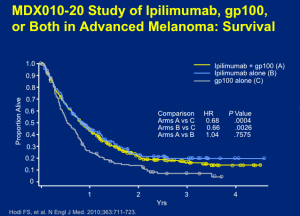
Screen Shot 2015-04-07 at 10.36.22 AM

Click to Enlarge


Ipilimumab was already a ten-year old therapy for which metastatic melonama patients were taking the drug for a few months course of treatment and who are still alive and well. Dr Flaherty states that we knew, when these results emerged, that there may be a truly, durable, lasting effect.

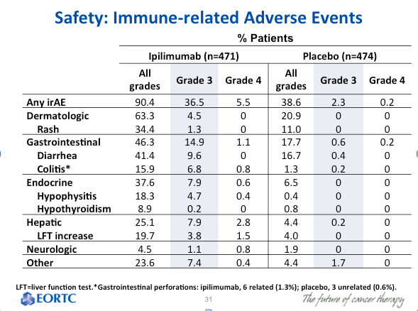
There is a cost to this type of treatment in terms of adverse events (AEs). All of the toxicities associated with this drug come from autoimmune attack(s) affecting the skin, large intestine, endocrine glands, or hepatic autoimmune injury. Why these tissues and not others? There are several theories, but not a firm understanding. If you focus on the ipilimumab monotherapy group, you can look at those who had severe autoimmune toxicity, i.e., you have to stop giving the drug and/or give high-dose corticosteroids to stop the autoimmune reaction. This occurs about 10 percent of the time. Everything short of this autoimmune toxicity appears, in short, in mild or moderate form and basically goes away without any immunologic intervention.
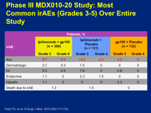
Phase III MDX010-20 Study:  Most Common irAEs (Grades 3-5) Over Entire Study

Click to Enlarge


The idea is that we think we’re unmasking normal tissue tolerance, but in a largely reversible way. About two thirds of patients who receive ipilimumab have some form of autoimmune toxicity, only about 10 percent have enough of it that it creates a truly life-threatening scenario for which we need to intervene. Not all of the toxicities manifest at the same time. Dr Flaherty feels that in major melanoma programs, we have become quite comfortable with regards to counseling patients as far as what to look out for and when to call with suspected reaction(s). Dr Flaherty believes that ipilimumab is a therapy that can absolutely be safely administered.

The other phase III trial that corroborated the effects of ipilimumab looked at patients taking ipilimumab (10mg/kg) plus decarbazine (850 mg/m2) or dacarbazine (850 mg/m2) plus placebo at weeks one, four, seven, and ten, followed by dacarbazine alone every three weeks through week 22. This trial was similarly positive with the addition of ipilimumab. The difference, as shown below, was really not very different from that of the ipilimumab versus vaccine trial.
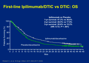
First-line Ipilimumamb/DTIC vs DTIC: OS

Click to Enlarge


The toxicities of the two-drug approach were modestly worse. When the FDA looked across the two data sets, they didn’t feel that the addition of chemotherapy was synergistic; therefore, they went with the previous study and approved ipilimumab as a monotherapy for metastatic melanoma.

In clinical practice, you can look at the pooled data set (below) and feel comfortable counseling patients that they have about a 20 percent chance of walking away with the diagnosis of metastatic melanoma by receiving this treatment (four doses over three months) with time-limited risk of toxicity.
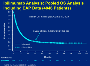
Ipilimumab Analysis:  Pooled OS Analysis

Click to Enlarge


We have learned that if you “cherry-pick” patients and only treat those with lower disease burden, the rate may be as high as 30 percent.

The PD-1 Pathway

What is the role of the PD-1 pathway in suppressing anti-tumor immunity? How might we think about deploying antibodies that try to intercept this negative immune regulator? We can target on either the PD-1 side or the PD-L1 side [with antibodies]. Scientifically, we would anticipate that this would alleviate the negative influence that the expression of this immune marker on tumor cells can produce in terms of silencing T-cells directly. There was a belief that PD-L1 target is actually safer than PD-1 targeting because you don’t disrupt PD-L2/PD-L1 interactions that a PD-1-blocking antibody would and there is some clinical evidence that this may be true.

Pembrolizumab

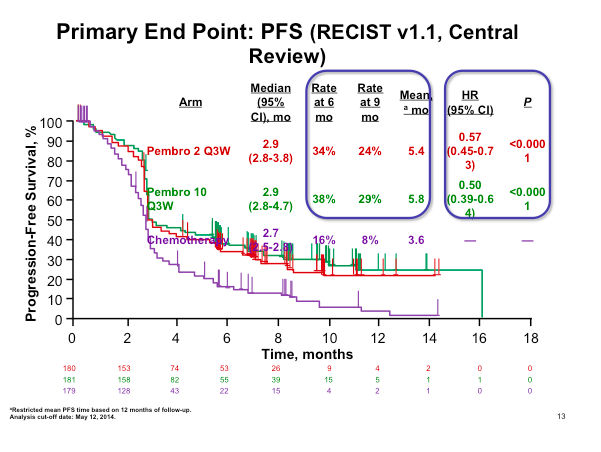
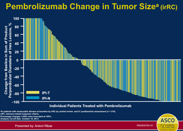
Pembrolizumab was FDA-approved in the late summer of 2014. In the pivotal, randomized study, patients who had received ipilimumab and/or a BRAF inhibitor, if they were BRAF mutant, were randomized to receive pembrolizumab 2mg/kg IV every three weeks (ongoing), pembrolizumab 10mg/kg IV every three weeks (ongoing) or chemotherapy. This was a trial that was aiming to show that patients who had exhausted the available therapies could benefit from treatment with pembrolizumab. This data set addresses the unmet need of patients who don’t get a benefit from ipilimumab.
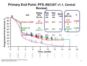
Primary End Point:  PFS (RECIST v1.1 Central Review)

Click to Enlarge


The data (above) is impressive when looking at pembrolizumab compared to chemotherapy at the level of progression-free survival. This is clearly a positive study and very important data for patients who had exhausted all of the other treatment options. It is true; however, that this therapy does not work for all patients and about half of patients will still experience disease progression. The number of patients who have objective responses, again these are patients who have not exhausted all other options, appears to be about 20-25 percent.

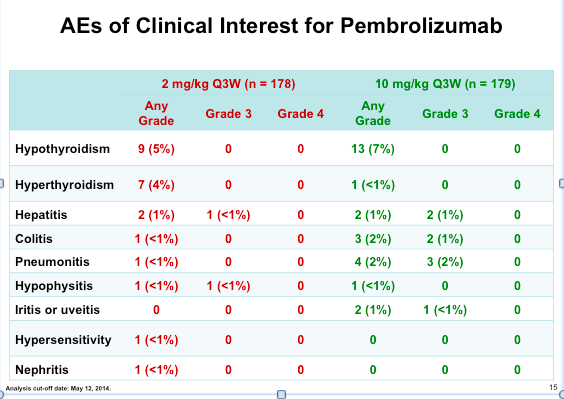
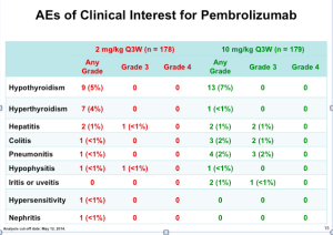
Two different doses were investigated in this study (2 mg/kg Q3W and 10mg/kg Q3W). Because there was no difference in efficacy, the lower dose, which is modestly safer, was the regimen that was approved. The toxicity is identical in type with ipilimumab. This type of immune therapy produces the same autoimmune reactions in terms of the affected organs and tissues. The issue is that the severity is clearly lower than that of iplimumab.

AEs of Clinical Interest for Pembrolizumab

Click to Enlarge


If you look at the Grade 3 and Grade 4 AEs (above), notice how there are only a few autoimmune toxicities in the approved pembrolizumab group (2mg/kg).

For our patients, this is a drug that can be effective and a bit less dangerous in terms of autoimmune toxicities.

Nivolumab

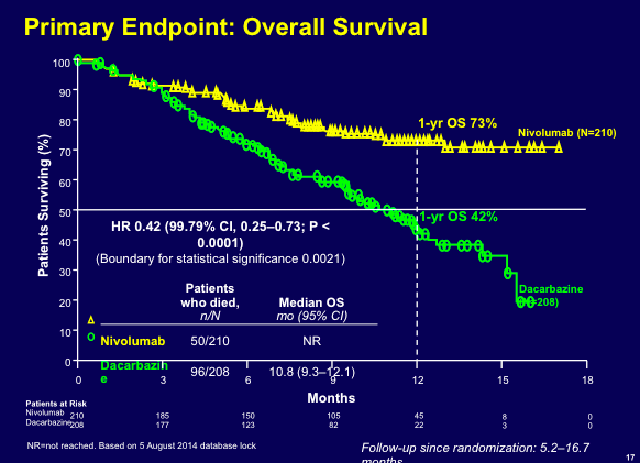
Nivolumab, another PD-1 antibody that was approved in December 2014, was studied in patients who had not received prior therapy. Patients were randomized to nivolumab (3 mg/kg IV every two weeks) plus placebo or placebo plus dacarbazine (1000 mg/m2 IV every three weeks). The thought here was that if you were going to conduct a clinical trial that was chemotherapy controlled, you couldn’t include BRAF-mutant patients because you wouldn’t offer them decarbazine as a front-line “cytotoxic” therapy. Patients in this study were stratified based on PD-L1 status.

This drug has a huge impact in terms of overall survival.

Primary Endpoint:  Overall Survival

Click to Enlarge


The data demonstrate a one-year overall survival rate of 73 percent. Many practitioners look at the evidence and would like to get PD-1 antibodies in the front-line. Currently, these drugs are approved for second- or third-line.

Progression-free survival clearly improved as well with nivolumab. These are immune therapies, but they have a clear impact on tumor burden as well. About sixty percent of patients have some demonstrable tumor effect.

When you see a response, the likelihood that the response is going to be durable through six and twelve months of follow-up is very likely.

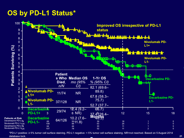
PD-L1 status is an interesting biomarker to which we should pay attention. The overall survival outcome for patients whose tumors express PD-L1 on their surface does better than the control group and the overall population. (see below)
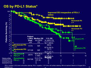
OS by PD-L1 Status*

Click to Enlarge


It is fairly clear that we can do some enrichment by using PD-L1 on the tumor as a biomarker.

Combination Therapy

In the medical oncology metastatic melanoma field, there is a lot of research around the concept of combining PD-1 and CTLA-4-blocking antibodies. Tumor responses in patients receiving nivolumab plus ipilimumab demonstrated that approximately 90 percent of patients responded after a follow-up of about 13 months.

Cli

There is still a group of patients who remain refractory, even to two-drug therapy. This phase I data ultimately launched a large phase III trial and we expect to see the data in June of this year (2015). The combination regimen cannot be given at the full dose of each drug; at least one of the therapies has to be attenuated. The data in the phase III trial is not quite as robust; however, we look forward to hearing the results in June.

When you look at the best available data with PD-1 monotherapy (pembrolizumab), this is actually not bad (below). Dr Flaherty feels that it is still unclear as to whether combination therapy is lifting us to a new plateau. We need more evidence to support that. Regarding safety, in patients who were taking combination therapy at the doses that were taken into the phase III trial, there was a sixty percent rate of severe (Grades 3 and 4) autoimmune toxicity. The results from the phase III data will help us to determine whether or not this combination therapy can be clinically useful.

Pembrolizumab Change in Tumor Size

Click to Enlarge


Adjuvant Therapy

There is some preliminary evidence on adjuvant therapy as we only have an interim result from a trial looking at ipilimumab monotherapy versus placebo in 951 patients with high-risk, stage III, completely resected melanoma. The primary endpoint was recurrence-free survival. If you focus on hazard ratio, there is about a 25 percent improvement. The question is whether or not this drug is really doing something more effective in the adjuvant setting than it would otherwise do in the overt metastatic setting. This is not clear—we need to see more data, more follow-up, and more overall survival evidence to know if this is a therapy that should be moved to the high-risk, resected setting. This therapy can have significant toxicity consequences, so we do have reason to be concerned.

Toxicities are substantial in the adjuvant setting. Notice the rate of Grade 3 and 4 toxicities. (below) This is the same drug that has a ten to twelve percent toxicity rate in the overt metastatic setting, but more in the adjuvant setting—partly because of the higher dose.

Safety:  Immune-related Adverse Events

Click to Enlarge


There were treatment-related deaths in this trial. This is not something that we can take lightly.

Vaccines

From 2010 to 2015, in the metastatic melanoma field, we have run about twelve phase III trials and they have all been positive, except for the most recent of the vaccine trials, a MAGE-3 peptide. Prior to this, we have had three peptide vaccine trials suggesting a detriment in overall survival. We still have not found a way to deploy these therapies. Dr Flaherty states that many of us are hopeful that either by using some biomarker selection strategy or, more likely, using these agents with immune checkpoint antibodies, we may be able to find utility in steering immune responses towards specific epitopes. Currently, there are no ongoing phase III trials.

Conclusions

Ipilimumab was the first-in-class immune checkpoint inhibitor and has demonstrated survival advantage. Long-term follow-up clearly supports survival impact with only three months of therapy. PD-1/PD-L1 is considered “best-in-class” based on higher response rates and better disease control; however, the percentage rate of long-term survivors is unknown. The combination of ipilimumab/nivolumab suggests synergistic toxicity and the question remains regarding synergistic efficacy. The adjuvant role of therapy is still not defined as we only have interim data for ipilimumab at the higher dose.


Dermoscopy: Clinical Pearls

Ashfaq Marghoob, MD

Dermoscopy Clinical Pearls:

  • The presence of leaf-like and spoke wheel-like structures seen with dermoscopy is 100% specific for the diagnosis of basal cell carcinoma.
  • The presence of vessels arranged in a string of pearls pattern seen under dermoscopy is 100% specific for the diagnosis of clear cell acanthoma.
  • The presence of white blotches and strands within the same lesion seen with polarized dermoscopy is highly suggestive of basal cell carcinoma
  • Angulated lines forming a zigzag pattern or forming polygonal shapes is suggestive of pigmented actinic keratosis or lentigo maligna
  • The presence of any of the 10 melanoma specific structures in a melanocytic lesion should prompt consideration for a biopsy

Cutaneous Oncology: Clinical Pearls

George Martin, MD & Ted Rosen, MD

Why field therapy?

Data from Europe using a variety of techniques (including full face cross-polarized light examination, fluorescence photography, high definition optical coherence topography and reflectance confocal microscopy) clearly validate the concept of a “field” of abnormality in close spatial proximity to visible AK lesions.

Facial AKs:

Although we did not mention this in our talk, for those of you who use 5-FU, please remember that the Phase III data on 0.5% 5-FU demonstrated that 1 week of daily use of 0.5% 5-FU cleared nearly 75% of individual AKs. Try: 0.5% 5-FU QD x 1 week, wait 1 month, then follow with 2 -3 weeks QD application to “clean up” remaining AKs. This regiment has gained widespread acceptance by patients and physicians as a more tolerable field therapy. 5% 5-FU BID is equivalent to 0.5% 5-FU and can be used interchangeably.

Ingenol mebutate 0.015% applied nightly x 3 has been a remarkably effective therapy with great patient compliance. However, it’s FDA approval was for limited areas (25 cm2). As a full-face therapy, it appears very effective; however, controlled studies on “full face” clearance/efficacy is pending. Patients need to be counseled that they will experience a moderate to severe “sunburn-like” effect beginning 4 hours after application. This is likely due to its MOA, which includes a direct cytotoxic effect. Analgesia is generally required. Clinically, we have observed while treating full-face, ingenol mebutate is selective for AKs with mild/moderate erythema between AK lesions.

New data, both short term (11 weeks) and long term (one year), support the benefit of using ingenol mebutate in combination with standard liquid nitrogen cryotherapy. Cryotherapy is performed first, and then ingenol mebutate used per approved protocol three weeks late. At one year, the combination therapy has a higher complete clearance rate than cryotherapy followed by vehicle control. Off label, ingenol mebutate appears promising for the management of actinic cheilitis in the three day, FDA-approved regimen. More studies are needed to validate this idea.

If you perform PDT in your practice, and use 1 -> 3-hour ALA incubation periods, recent Phase II studies from DUSA show that 1, 2 or 3 hour incubation periods are roughly equally efficacious (35% – 50% clearance) but 2 treatments 8 weeks apart are required for most patients to achieve >70% individual lesion clearance. To maximize the efficacy of a 1-hour incubation, consider pre-treating with 5-FU for 1 week to the face or 10 days to the scalp then perform a 1 hr. ALA incubation. This combination will eliminate the need for a 2nd PDT. For those patients with refractory facial AKs, consider pretreating for 7 days with 3.75% imiquimod followed by ALA PDT (1-3 hour incubation). Excellent long-term results (18 months) have been observed when destructive techniques such as PDT are combined with immune modulators.

Can ALA PDT be “painless”?? Try incubating for 15 mins with ALA and then place the patient under the blue light for 1 hour. Preliminary results (G.Martin MD) demonstrate that ALA PDT as monotherapy or in combination with 5-FU or 3.75% imiquimod, employing this technique is in fact “painless”. Individual AK clearance rates of 50% were demonstrated in a proof of concept small patient study. Large-scale studies are warranted to determine efficacy. Network meta-analysis (comparing different techniques against one-another) suggest that PDT is the optimal field therapy for AKs.

The focus of PDT has been aimed at treating AKs. Is there evidence that PDT may prevent BCCs? Data from studies on basal-cell nevus syndrome patients demonstrate long-term clearance and prevention of new BCC development compared to non-treated areas of the trunk following ALA PDT using red light.

The use of 3.75% imiquimod for diffuse facial AKs while effective, results in substantial “downtime” of nearly 6 weeks. Consider 3.75% imiquimod QD x 7 days, 2 weeks rest, followed by once weekly applications. There will be some initial unsightliness during the 1 week of continuous use. Individual AKs are seen to clear with continuous application with minimal irritation. Chronic immune stimulation (>1 year) appears to be helpful in limiting AK recurrences and may prove over time to inhibit the development of invasive SCC. Large-scale studies are warranted.

Melanoma Clinic: Lunch, Lesions, and Lessons: Part 4

Hensin Tsao, MD, PhD

Ilona Frieden, MD

Philip LeBoit, MD

Keith Flaherty, MD

Case Study

An 11 year-old boy presents to your office with his parents for the evaluation of a pigmented band on the right thumb. What would you do next?

Screen Shot 2014-12-05 at 2.46.54 PM

  1. Photo and follow up in 6 months
  2. Reassure and continue careful surveillance at home
  3. Perform an matrix biopsy with local anesthesia
  4. Matrix biopsy under general anesthesia

Melanonychia Management

Melanonychia is not rare; however, nail melanoma in children is rare. The pre-test probability of a benign outcome is high. Most of the time we can photograph and follow the patient over time. In an unscientific perusal of cases at UCSF, Dr Frieden found that the majority of the patients were of Asian descent. Occasionally, a biopsy is performed. In a 20-year cohort, there were no melanoma deaths in childhood from nail melanoma.

Special Sites and Concepts of “Mino”

Special sites, such as the nail, scalp, oral and genitalia, require careful scrutiny and some pathologists appear to have a better handle regarding the appearance of melanocytic lesions. The usual rules, both clinically and pathologically, often times do not apply. Definite cases of nail melanoma in childhood have been reported and there have been issues in the pathology of nail melanocytic biopsies. Richert and colleagues conducted a retrospective cohort of 30 biopsied lesions in Belgium. Seven out of the 30 patients were age 20 or younger and none of them had melanoma. (Richert B, et al. J Am Acad Dermatol. 2013;69(1):96-140.) A 2008 review article reported two cases of nail melanoma in children with skin type 3 and 4 in a 14 year-old and a 6 year-old. Both were diagnosed with having melanoma in situ (MIS) with onset of melanonychia less than one year of age. There were eight other reported cases in the literature with six diagnosed as MIS. Two had lymph node disease and neither presented as melanonychia. (Iorrizzo et al. Dermatol Surg. 2008;34:974-8)

“Struck by lightening versus severe bike accident?” Dr Frieden thinks about nail melanoma in terms of risk assessment. In general, we would not worry about being struck by lightening. It doesn’t mean it doesn’t happen, but it is extraordinarily rare.


Melanoma Clinic: Lunch, Lesions, and Lessons: Part 3

Hensin Tsao, MD, PhD

Ilona Frieden

Philip LeBoit, MD

Keith Flaherty, MD

Case Study

A forty year-old woman presents to her PCP with a lesion that she had removed near her left knee (cryotherapy) about one year ago. She thinks something is returning. “It is a tiny miniscule nodule that appears to be a scar.” This appears to be a dermatological issue and “she wants to see her old dermatologist to have it rechecked.” What is an appropriate next step for the physician?

  1. Refer and document
  2. Refer and follow-up with phone call in 1 m
  3. Refer and call the local dermatologist
  4. Refer and follow up with letters until lesion is removed

If you have a general concern, three certified letters can be a good approach. It is extremely important to follow-through and maintain good communication so that you are not held liable.

Seven months later, the patient returns to the PCP with her parents. She was diagnosed with an invasive melanoma. She didn’t see the dermatologist, whom she was supposed to see last spring, until just recently. The patient was referred to Massachusetts General Hospital.

Melanoma on the left knee showed NMM; 7.5mm thick, ulceration 0.4mm, 12 mits/mm2; no microsatellites. She underwent wide resection and sentinel lymph node biopsy (0/8). One year after the diagnosis, she returns for a follow-up and informs you that she wants to become pregnant. She is now 41 years old and she had a miscarriage several years ago. This scenario is not uncommon and Dr Tsao has had referrals regarding this situation.

What do you tell a 41 year-old woman with a stage IIc (life expectancy 30-50%) melanoma about pregnancy?

  1. Fine to become pregnant
  2. Wait at least 2 years
  3. Wait at least 5 years
  4. Wait at least 10 years

Dr Flaherty comments that the fundamental question that we don’t have a great answer to is whether or not every instance of microscopic metastatic disease would ultimately manifest as overt metastatic disease with time. Or, are there patients who can live chronically, i.e., forever with microscopic disease? We do know of late relapses with melanoma. Dr Flaherty tends to tell patients the suggestion that microscopic metastatic disease will convert to macroscopic metastatic disease and the worst that could happen, in the pregnancy setting, is that you could influence the conversion, in other words, it could happen sooner. It is also important to discuss where patients are on the risk curve. The risk of occurrence falls with time.

The risk of relapse after 10 years is actually not clear because high-risk lesions should have relapsed by then while thin relapses continue to emerge. There are differences in the kinetics of relapse between a thin melanoma and a thick melanoma. The bigger the tumor, the more aggressive it is. If the patient survives to year five or ten, it is less likely that something will happen. In fact, there might be a cross over point, probably around year 10, whereby if you had a thick tumor and you’re still alive, the likelihood of it recurring is quite low.

Pregnancy and Melanoma

There are many case control series of melanoma during pregnancy versus not during pregnancy. A 2008 study found no evidence that pregnancy worsens the prognosis. (Cancer Causes Control. 2008;5:437) Some studies suggest that melanoma during pregnancy may be slightly thicker but survival is not altered that much.

The patient visits with a high-risk obstetrician who clears her to become pregnant. Three months later, she finds out that she is pregnant. How do you follow a pregnant person with a history of melanoma?

  1. Routine post-melanoma surveillance
  2. Routine surveillance with total body photography
  3. Increased surveillance every three months
  4. Increased surveillance with total body photography

Total body photography could be a viable option. There is really no literature on dermoscopy in pregnancy. These patients should be followed very carefully. Regarding post-melanoma surveillance, you don’t need to necessarily change your practice because of pregnancy. There are no great studies that convincingly demonstrate a higher risk of relapse during pregnancy. This is highly anecdotal, yet it is very compelling and memorable when it happens.

At week 17 of her pregnancy, she noticed a small, firm nodule within her melanoma scar. Wedge biopsy showed recurrence of melanoma with neurotropism. Wide resection cleared recurrence and a skin graft was placed.

Management of Melanoma Recurrence During Pregnancy

There are treatment options; however, there are no data for metastatic disease during pregnancy. With regards to immunotherapy, high-dose IL-2, which is a physiologic stress, poses a theoretical risk of abruption (in the setting of hypotension). Ipilimumab is likely the first-line choice. Looking at targeted therapy, BRAF inhibitors showed no risk of teratogenicity in limited preclinical studies and MEK inhibitors are theoretically of greater concern; however, that may not be the case in combination with BRAF inhibitors. Chemotherapy is rarely an attractive first-line option in any case.

Package Inserts

  • Vemurafenib
    • Pregnancy Category D
    • Vemurafenib can cause fetal harm when administered to a pregnant woman based on its mechanism of action
  • Iplimumab
    • Pregnancy Category C
    • Use ipilimumab during pregnancy only if the potential benefit justifies the potential risk to the fetus.

The patient received XRT to the left knee graft area with shielding of the fetus to prevent radiation crossover. At 36 weeks, she underwent a Caesarian section. A histological examination of the placenta did not reveal melanoma. (There have been reports of metastases to the placenta or fetus) One month after delivery she noticed two subcutaneous nodules in the mid-calf distal to the scar site. A biopsy showed metastatic melanoma consistent with in-transit disease. PET/CT showed multiple left lower leg FDG avid nodules, but no visceral metastases. Both radiologically and clinically, she’s beginning to develop at a faster pace.

What would you recommend now?

  1. Isolated limb perfusion
  2. Targeted removal of individual lesions
  3. Genotyping with anti-BRAF agent if BRAF+
  4. Systemic treatment with IL-2 or immune checkpoint antibody

In-Transit Metastases

We lack high-level evidence for the optimal approach in this case. According to the NCCN guidelines, enrollment in a clinical trial is the preferred approach. Options for local therapy include complete surgical excision, intralesional injection (BCG, IFN, IL-2), local ablation therapy, topical imiquimod for superficial dermal lesions, and consideration of palliative XRT for unresectable disease. Isolated limb infusions/perfusions with melphalan could be considered as a regional therapy option as well as the use of systemic therapy. Systemic used to be considered as bottom of the list; however, it is being utilized more and more.

The issue with regional therapy is that it has no impact on disease elsewhere. With regards to intralesional therapy, Weide and colleagues found a high response rate after intratumoral treatment with IL-2 in a phase two study of 51 patients with metastatic melanoma. The 2-year survival rate was 77 percent for patients with stage IIIB/IIIC disease. Efficacy and survival did not differ between patients who had greater than or equal to 20 lesions and patients who had less than 20 lesions. (Weide B, et al. Cancer. 2010;116(17):4139-4146.)

Alexander and colleagues analyzed factors that influenced the outcome in patients with in-transit malignant melanoma undergoing isolated limb perfusion using modern treatment parameters. Between May 1992 and February 2005, 91 patients underwent a 90-minute hyperthermic ILP. There were 62 complete responses (69%) and 23 partial responses (26%) in 90 assessable patients.

The patient underwent isolated limb perfusion with melphalan but unfortunately continued to progress. She continued to progress through multiple experimental treatments over the ensuing year and she expired six years after her initial diagnosis.

Pregnancy and Melanoma-Take Home Points

  • Pregnancy does not appear to compromise survival
    • “Wait time” considerations include:
      • Potential treatment complications during pregnancy
      • Age of patient and desire for children
      • Personal outlook (e.g. religion, “abandoning” children)
      • Availability of oocyte cryopreservation
    • Risk extinction is more obvious for thicker tumors
  • Bottom line (not scientifically based)
    • For patients with multiple DN- watch q3m’s during pregnancy
    • For MIS and thin melanomas- OK to start (need discussion)
    • For melanomas >1mm- wait 2 yrs unless age is major concern
  • Discussion (Push/pull model)
  • Push information-
    • There is the chance of relapse and death (estimate prognosis)
    • Come to an agreement on a course of action
    • There is a tiny chance of transplacental metastasis
    • Pregnancy per se does not appear to influence outcome- gestation cannot be an instrument of guilt or regret
    • Need partnership with high-risk OB and possibly early consult with medical oncology
    • Patients should probably be monitored more frequently
    • Inform patient about possibility of additional biopsies during pregnancy
    • Inform patient about oocyte cryopreservation
  • Pull information (warning: takes time and may be difficult emotionally)-
    • Does the patient understand the implications of relapse during pregnancy (including the possibility of termination)?
    • What are the plans should baby survive but mother does not?
      • Discussion becomes more concrete and intense as relapse risk increases
      • Wait time is meant to clarify this risk a bit
      • May need social work to help through these conversations


MauiDerm News Editor-Judy Seraphine