Lasers: Red vs Blue in PDT

Its Blue

A study reported late last year was carried out to determine whether prior microneedling would enhance the penetration of topical aminolevulinic acid HCl (Levulan) and thus enhance photodynamic therapy (PDT) and possibly also result in a better cosmetic outcome vs PDT alone in patients with AKs.

In this trial, 20 patients each with ≥4 non-hyperkeratotic AKs on each side of their faces were randomized to receive multiple passes with a microneedling device on one side of their faces, followed by application of aminolevulinic acid HCl to the entire face.

The aminolevulinic acid HCl was allowed to incubate for 1 hour and this was followed by exposure to blue light (Blu U) for 1000 seconds.

For the 19 patients who completed 4 months of follow-up, the mean AK reduction on the microneedling side was 89.3% vs 69.5% on the PDT alone side. In addition, 13 of the 19 patients had a noticeable improved cosmetic appearance on the microneedled side of the face (Spencer, 2016).

No, Its Red

Aminolevulinic acid hydrochloride (BF-200 ALA, Ameluz) was recently approved by the United States Food and Drug Administration for PDT using the BF-RhodoLED lamp, a narrowband, red light illumination source, for lesion-directed and field-directed treatment of AKs of mild-to-moderate severity on the face and scalp.

This approval was based on results from 779 patients with 4-8 mild to moderate AK lesions. The results obtained from these studies demonstrated that BF-200 ALA was significantly superior to the standard of care, with a complete patient response rate of 91% when paired with BF-RhodoLED PDT lamp (CenterWatch, 2017).

In addition, BF-200 ALA showed positive long-term effects with low recurrence over the course of 12 months (Dirschka, 2013).

In a pivotal phase III trial performed on entire treatment fields, BF-200 ALA demonstrated long-lasting skin rejuvenation effects in sun-damaged, but asymptomatic, skin regions (CenterWatch, 2017; Reinhold, 2016).


References

CenterWatch. Ameluz (aminolevulinic acid hydrochloride). 2017. Available at: http://www.centerwatch.com/drug-information/fda-approved-drugs/drug/100150/ameluz-aminolevulinic-acid-hydrochloride

Dirschka T, Radny P, Dominicus R, et al. Long-term (6 and 12 months) follow-up of two prospective, randomized, controlled phase III trials of photodynamic therapy with BF-200 ALA and methyl aminolaevulinate for the treatment of actinic keratosis. Br J Dermatol. 2013;168:825-36.

Reinhold U, Dirschka T, Ostendorf R, et al. A randomized, double-blind, phase III, multicentre study to evaluate the safety and efficacy of BF-200 ALA (Ameluz(®) ) vs. placebo in the field-directed treatment of mild-to-moderate actinic keratosis with photodynamic therapy (PDT) when using the BF-RhodoLED lamp. Br J Dermatol. 2016;175:696-705.

Spencer JM, Freeman SA. Microneedling Prior to Levulan PDT for the Treatment of Actinic Keratoses: A Split-Face, Blinded Trial. J Drugs Dermatol. 2016;15:1072-4.

Advances in Treating Varicose Veins and Telangiectatic Leg Veins

Mitchel P. Goldman, MD

Dr Goldman is a Volunteer Clinical Professor of Dermatology at the University of California in San Diego, Immediate Past-President of the American Society for Dermatologic Surgery (ASDS), and an expert in Sclerotherapy publishing 5 textbooks and dozens of medical papers on varicose and telangiectatic leg veins. In this presentation, he provides us with an update on the treatment of varicose and telangiectatic leg veins.

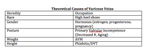
Thirty percent of the population will have varicose and telangiectatic leg veins by age twenty. As we age, the incidence increases, i.e., 80 percent of the population will have varicose and telangiectatic leg veins by age 80. There are many reasons for developing varicose veins. The incidence among men and women is about equal. It is important to remember that up to fifty percent of varicose vein patients will develop DVT, SVT, and/or ulceration in their lifetime.

Dr Goldman feels that the more factors you have, the more likely you are to get varicose veins.

Screen Shot 2014-12-26 at 7.47.31 AM

When you look at longitudinal studies of large populations (Bochum Study—Schultz-Ehrenberg, 2004), you will see that the veins start out as spider veins and then gradually, the older you get the more reticular or truncal varicose veins appear.

Screen Shot 2014-12-26 at 7.47.20 AM

Why do people come to us for their varicose veins? They come primarily not because of symptomatology, but for cosmetic reasons. Most physicians think that veins are just a cosmetic procedure and not a real medical problem, but this is an actual medical condition. In Dr Goldman’s office, even when leg pain is not a presenting concern, it is not uncommon for patients to tell him that their legs feel better after treatment.

Varicose and Telangiectatic Leg Veins: Symptoms

  • Heaviness
  • Aches and Pains
  • Night cramps/Restless legs
  • Ankle edema

Why is there pain? There’s pain because the venous hypertension, i.e., veins that you can see on the surface, are actually surrounding and pressing on nerves. Venous reflux can cause varicose veins, skin changes, skin ulcers and swollen legs. This is why dermatology is the specialty that should be treating this condition. In Europe, it’s the dermatologist who is the phlebologist.

Current treatment options include:

  • Compression stockings
  • Sclerotherapy
  • Phlebectomy
  • Stripping and ligation
  • Endovenous laser
  • Endovenous radiofrequency

When treating patients with varicose veins, insurance companies will first ask you if the patient has failed compression stockings—implying that the patients should be wearing these stockings for the rest of their life. If patients are complaining of pain, an easy way to see if it’s caused by the veins is to put your patients in graduated compression stockings. If the pain goes away, then you know that sclerotherapy or treatment of the veins is going to make the pain go away as well.

A study by Schul et al compared compression versus sclerotherapy for patients with isolated refluxing reticular veins and telangiectasia. They looked at 50 women with symptomatic veins with normal saphenous and deep veins. Ninety percent of the women required OTC analgesics. The subjects were randomized to thigh high 20 to 30 mmHg graduated compression for six weeks versus sclerotherapy. All compression patients crossed over to sclerotherapy. The researchers found that graduated compression improved aching, pain, cramps, and restlessness and sclerotherapy improved everything plus made the veins go away. (Scul et al. Phlebology. 2011;26:148.)

When you treat varicose or telangiectatic leg veins, you have to be logical and do it in an order. You can’t just hit or miss with vein injections and expect a good result. It would be the same analogy as if a patient had a skin cancer on the face—you would not just treat little bits here and there.

A Logical Progression

  • Begin with cut-off of reflux from SFJ/SPJ
    • Endoluminal RF or LASER Closure or
    • Foam Sclerotherapy
  • Ambulatory phlebectomy or Foam Sclerotherapy of veins > 4mm
  • Sclerotherapy of remaining veins
  • Then consider LASER or IPL

When a patient presents with telangiectasias in one little area, you need to remember that telangiectasias are fed by deeper reticular veins all of the way into the deep venous system. Everything is connected. When Dr Goldman treats a patient, he doesn’t do a section at a time, nor does he bill by the amount of solution that he’s using. He treats per leg. It is imperative that you treat the entire venous system in one leg in order to minimize the number of treatments.

Screen Shot 2014-12-26 at 7.48.10 AM

Screen Shot 2014-12-26 at 7.48.33 AM

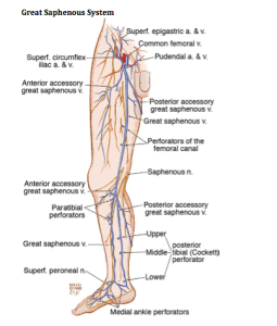
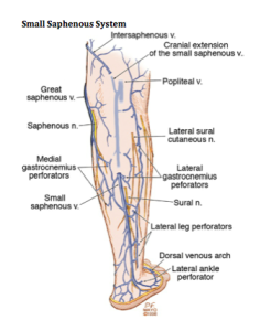
What gets us into trouble when we don’t know the anatomy is that we have to figure out where the veins are coming from. There are times when the veins don’t come from the incompetent great saphenous vein. Dr Goldman uses a duplex machine to determine where the veins are coming from.

As Dermatologists, we tend to see the lateral subdermal plexus. When patients present with little telangiectasias on the lateral aspect of the thigh, which is very common, we must realize that it’s all coming from an incompetent reticular vein. You must treat the entire leg.

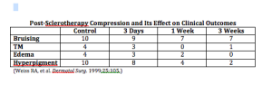
If you leave a vessel open after it’s been injured, your body is going to reconstitute it and the endothelial cells are going to keep trying to reestablish the vessel. If you compress the vessel after you’ve injured it, you can decrease the extent of thrombosis, which decreases the risk of recanalization and pigmentation allowing for the vessels to go away quickly. Walking is another way to do compression. When you walk, you’re basically siphoning off the blood flow from the superficial venous system into the deep venous system and that’s doing a physiologic form of compression. You want to minimize the thrombosis as best as you can.

Screen Shot 2014-12-26 at 7.48.44 AM

You want to have patients who are going to follow your directions as compression really does improve outcomes. Dr Goldman only uses thigh-high stockings for the patients. He stocks them in his office because the pharmacies and medical supply offices do not always know how to best measure for stockings correctly. The stockings must be applied immediately after treatment.

What is sclerotherapy?

Sclerotherapy is a controlled thrombophlebitic reaction that began hundreds of years ago. There are two solutions approved by the FDA that can be used—sodium tretadecyl sulfate (STS) and polidocanol (POL). Glycerin is also an approved solution.

Sclerosing Solution Matched to Vessel Size

  • Telangiectasias < 1mm in diameter
    • Very small volumes of hypertonic saline and/or dextrose
    • 0.1% – 0.25% STS liquid
    • 0.25% – 0.5% POL liquid
    • 72% Glycerin mixed 2:1 with 1% lidocaine +/- epi
  • Reticular veins > 1mm in diameter
    • 0.25% – 0.5% STS foam
    • 0.5% – 1.0% Pol foam

When you start injecting a vein, you have to go back to your high school physics. You have to fully inject until you stop seeing solution in the vein because once you stop seeing where the solution is flowing, it’s flowing deep—where you don’t particularly want it.


  • Volume of a vein-Vv=ttD2/4L (D= internal diameter and L = length)
  • 2ml into a 5mm travels 10cm; 0.5ml into a 0.2mm vein travels 6cm

Detergent solutions (sodium tetradecyl sulfate and polidocanol) are very efficient. They strip off the proteins from the endothelial cell causing its destruction.

Screen Shot 2014-12-26 at 7.48.54 AM

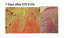
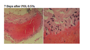
You can see, from the biopsies above, that there is a total wipe out of the endothelial cells. Using the same dosage of polidocanol, we don’t see the wipe out of the telangiectasia, it basically just attenuated the endothelium.

Screen Shot 2014-12-26 at 7.49.05 AM

When you’re using STS versus POL, the concentration is essentially two to one. POL is one half of the potency of STS. Both solutions are very comparable in efficacy.

Parsi and colleagues studied the in vitro effects of detergent sclerosants on antithrombotic mechanisms. They found that STS is antithrombolytic and does not destroy plasminogen. Reinjection of STS into a sclerosed vein can accelerate recannalization. POL has no effect on coagulation factors. If the sclerosant does not destroy the endothelium, it will stimulate inflammation, which produces t-PA with fibrinolysis. Parsi, et al. Eur J Endovas Surg. 2009)

Dr Goldman does NOT use hypertonic solutions for a few reasons. Osmotic agents like HS hurt and they only act within their osmotic gradient leading to a much higher risk of recurrence as well as pigmentation and pain.

Glycerin, in and of itself, is a hypertonic solution, but it is very safe. Dr Goldman has never seen pigmentation or ulceration from it. It is available and can be compounded from a USP pharmaceutical grade glycerin.

Screen Shot 2014-12-26 at 7.49.18 AM

Glycerin has comparable efficacy with less adverse effects. Dr Goldman usually adds lidocaine to the glycerin; the lidocaine does not affect the efficacy of the glycerin; however, it does minimize the pain.

Compounding Solutions

Compounding solutions are not to be done in sclerotherapy. This can open up an amazing amount of lawsuits that will not be defended by your malpractice.   Why does Dr Goldman use compounded glycerin? It has proven to be very safe and is basically the same product that neurosurgeons are using in the brain for cerebral edema.

Foaming

There are advantages to foamed sclerosants. They are increased in volume but the total sclerosant injected is decreased. The highest concentration contacts vessel walls causing total obliteration of the vessel. There is a very slow washout and long contact with the intima. The only problem with foaming is that one third of the population has patent foramen ovale causing the foam to go into the arterial system. It is very safe to foam and it is safe to have nitrogen bubbles in your body. Dr Goldman has been using foam for twenty years and has only had two patients complain of a headache or slight loss of vision for just a few seconds.

Conclusions

You really have to look at your patients and evaluate the results. Taking photographs is very important. It is important to have good communication with your patients in order to set realistic expectations.

Patient Preparation

  • Eat before hand to avoid vasovagal response
  • Don’t shave legs or use moisturizers day of treatment
  • Wear shorts during treatment sessions
  • Bring pre-fitted GCS to visit and FU
  • Patient is recumbent, ideally on power table

Equipment and Supplies

  • Good light   vein light   polarizing light
  • Comfortable table
  • Comfortable chair for you
  • Digital Camera
  • Sclerosing agents
  • Normal Saline
  • Alcohol soaked cotton balls
  • 4×4 Gauze sponges
  • Graduated compression Stockings
  • Needles
  • Syringes
  • (3 and 5ml + 2 way connector)
  • Mayo Stand
  • Hazardous waste container
  • Gloves
  • Chucks

In thirty years, Dr Goldman has never had major problem; however, allergic reactions and anaphylaxis can occur, so you must be prepared with resuscitation equipment. You should have an office protocol in order to deal with emergency situations.

Injection Technique for Telangiectasias

  • Use 30 gauge needles ½ inch long
  • It usually takes 0.1-0.2 cc’s to inject a spider vein
  • Avoid forceful pressure
  • May bend needle – 30 degrees
    • 30 gauge, plastic hub disposable
    • 33 gauge dull quickly, bend too easily, not disposable
  • Can’t draw blood back, and not necessary
  • Inject very slowly, with little pressure, until blood cleared
  • Graduated compression hose (30-40 mm Hg) for 1 week
    • Reduces pigmentation
    • Better response
  • Identify patterns and groups, e.g. lateral venous plexus
    • Use additional tricks if necessary
  • Begin injections at logical starting points (reflux sites) or base of telangiectatic web
  • Keep your eye on injection site at all times
  • Minimize volume of solution per site
  • Recognize earliest bleb formation and stop!
  • Use minimal sclerosant concentration

Again, the most important thing is that you do this logically. You want to eliminate the high-pressure reflux first. Treat the small varicose and reticular veins initially—associated telangiectasias should be treated on the same day. You want to treat from proximal to distal and treat the entire length of the vein in one session. Dr Goldman follows up with the patient six months post-procedure for maintenance.


Zappin the Tats

Suzanne Kilmer, MD

At MauiDerm 2014, Dr Kilmer, an expert in lasers and light devices for dermatological procedures, discusses tattoo removal…

For tattoo removal, we generally use the visible spectrum of light.  Years ago, we used CO2 laser or salabrasion for tattoo removal. The use of the CW laser resulted in trans-epidemal loss of ink, significant thermal damage and frequent scarring. The newer Q-Switched Lasers shatters the ink into smaller particles then utilizes macrophages to remove the ink; there is some trans-epidermal loss, yet, there is rarely any scarring.

There are several issues to assess regarding tattoo removal. Pulse duration can be performed in either the nanosecond domain or more recently, the picosecond domain; the picosecond domain may be better for breaking up the ink and it is and may not be as wavelength dependent. Remember that longer wavelengths penetrate deeper and, very importantly, is the fact that the ink’s absorption determines the best wavelength. Fluence/spot size should also be considered. We want the largest spot size; however, we need sufficient fluence. Larger spot sizes allow deeper penetration of effective fluence as long as the laser has sufficient power. Regarding treatment intervals, six to eight weeks is generally the best timing. Of note, dark tattoos are much more easy to treat versus multiple colored tattoos. Green ink responds best to red light and red ink absorbs more in the green light.

Some of the side effects with tattoo removal include ink darkening, incomplete removal of ink, allergic reactions and infections.

Tattoo Treatment Improvements

Over 25 years ago, we went from long pulse to QS laser treatment. QS treatment demonstrated a dramatic improvement in ink reduction and side effect profile. This was the first time that we were able to remove tattoos without scarring. We started with 694nm then added 1064/532 and then 755nm wavelengths in order to improve color removal. This was performed for over 20 years with minimal improvement. In the last two years, we have shortened the pulse width to long pico/ultrashort nanosecond domain. Some studies demonstrated that multiple treatments in the same day increased ink clearance.

Picosecond lasers have a greater photoacoustic effect. In 2012, Cynosure came out with the first picosecond laser and it clearly demonstrated an increased clearance of ink when compared to the QS laser.

Picosecond lasers can also improve ink clearance in resistant tattoos with just two treatments when compared to QS lasers which showed no change over seven treatments.

Another company, Cutera, is looking at using other wavelengths with a novel picosecond laser. They are looking at a 1064nm with 532 component for picosecond domain.

Kossida & Anderson first reported faster tattoo clearing with single versus four treatments on the same day. After treatment, you should wait 20 minutes for whitening to clear “R20”. The photoacoustic effect and shattering of ink particles creates a cavity and as nitrogen gas flows in, you can see whitening. Nitrogen is slow to dissolve out (hence prolonged whitening). A study by Kilmer and Ibrahimi supported repetitive treatment on the same day in order to expedite tattoo clearance. They explored effectiveness of one versus two versus three versus four treatments (to alleviate scheduling nightmares.)  They found that two treatments were better than one but then they saw a declining efficacy with further treatment. Geronemus et al used an optical clearing agent, PFD, eliminating the 20-minute interval to clear whitening.

R20 Results-QS Laser

We have found that multiple treatments are better than single treatment on the same day for most tattoos. There is less of a difference between two, three and four treatments on the same day. Increased swelling may occur, as noted by patients and the treating nurse. There was no increase in pain; in fact, most patients felt less pain with subsequent treatment. We also saw no difference in PIPA and no scarring was noted. Generally, all patients preferred a more rapid tattoo clearance.

In summary, ultrashort pulse widths are better, but not always and multi pass is better, usually.


MauiDerm News Editor-Judy Seraphine


Introduction to Lasers and Light (And a Touch of RF and Microwave)

E.V. Ross, MD

Dr Ross, a leader in the field of laser and light therapy, provided the audience with an in-depth overview on lasers and light. Dr Ross begins by reminding us that we tend to look for the “easy” way out to make our patients look their best with the least downtime. In order to be efficient and effective for his patients, Dr Ross likes to have many lasers in one room; therefore, providing the best possible technology.

It is important that we really understand laser physics. You need to know some math, but it doesn’t have to be difficult math. You can take very complex mathematical relationships and break them down into very simple algebraic relationships. By doing so, you can actually apply your lasers and other technologies quite confidently. It’s imperative to understand how they work because if you don’t, you can get into trouble very fast. Remember to look, listen and feel….listen to the reaction, look at the laser, look at the patient at every pulse to be sure the endpoint is what you want to see because ultimately, the endpoint is more important than the physics.

When we talk about lasers, we need to know some basic definitions. One of the most important terms is fluence; fluence is simply the light dose, i.e., the amount of energy that we are investing per surface area for a particular application.  We used to speak a lot about power, e.g. 7 watts power or 12 watts power. Currently, we don’t talk much about power because most of our lasers today are pulsed lasers. What really matters are wavelength, fluence and pulse duration. Pulse Width is  very important, i.e., the time over which energy is delivered. Spot size, which contributes to the intensity inside the skin, is mainly important for visible light lasers and infrared lasers because a larger spot will penetrate better.

LASER, Light Amplification by the Stimulated Emission of Radiation, is a concept that Einstein predicted back in the 1920s, but it was not realized until May of 1960.

Why is laser different than a lot of other light sources? We can use a lot of non-laser light sources in dermatology and certainly achieve nice results. Intense pulsed light is a great example. There are some features of lasers that make them helpful for dermatology and helpful for certain applications, but not always necessary for every application.  (See http://www.colorado.edu/physics/2000/lasers/index.html)

Why is it important to differentiate laser light from non-laser light? Dr Ross explains that it is because of engineering more than anything else. Most of our targets that we treat in dermatology have multiple wavelengths as far as their absorption. Lasers are a convenient way to deliver light, i.e., deliver photons to the target. This is why lasers are so popular. You can put lasers into a fiber, you can have monochromatic light, and you can deliver very high power. It’s the only way to really deliver very short pulses in nanoseconds and picoseconds for certain applications.

Why do we need to know how/why lasers, etc. work?

Dr Ross states that one important reason is that when your laser breaks down, which can happen, you want to understand why it isn’t working. If you have a good understanding of how your laser works, you may be able to correctly diagnose that. If you see that your laser isn’t working right, often times there’s an error code. It’s important that you write down the code and report that to the technician. Sometimes it’s as simple as restarting the system.

You should be able to troubleshoot laser problems in a logical way…Device malfunction is one problem, an example is a temperature sensor malfunction. However, a bigger problem could be your lack of familiarity with the device; most laser problems are caused by the operator because they’re not familiar with how the device works—sometimes they are rented and sometimes they are only used once per week. Other factors that contribute to laser problems include poor patient selection, operational errors, poor post-treatment care, and simply bad luck.

Dr Ross reminds us that we have to think. Most of the time when we get into trouble with lasers it’s because we weren’t thinking—invest all of your brain power into that particular case while you’re performing the procedure and that means paying particular attention to endpoints.

Four years ago right before the Christmas holiday, Dr Ross had a typical case of a woman who came in for treatment of telangiectasias with a pulsed-dye laser. He used the pulsed-dye laser, 10mm spot, 7 J/cm2, and a 10-millisecond pulse and everything was looking fine. The patient developed some mild purpura and she was incredibly upset. So what happened? She didn’t have any blisters and everything else was fine. Dr Ross went back and looked at the beam profile and found that it was off by about 2mm (8mm instead of 10mm).  What had then happened was that the fiber was damaged so they were delivering the right energy but in a smaller spot; therefore, causing the fluence to become higher and the purpura threshold had become breached. The technicians then fixed the beam profile and there was no more purpura. Dr Ross reminds us that sometimes we may have to do some detective work to figure out what’s going on.

Broken mirrors can also compromise your laser experience.  Particularly with the CO2 laser, if your beam profile doesn’t look good it may be due to a broken mirror.

When your calibration doesn’t pass,  the most common reason is because of a bad lamp.

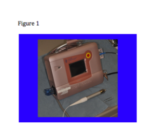
What we’re seeing now is a progressive change in laser technology. Lasers are becoming quieter, cooler, smaller, better and more reliable. We now have small, portable lasers (see figure 1) and maybe, in the near future, we’ll see small lasers that can treat everything from tattoos to resurfacing.
Screen Shot 2014-05-26 at 7.50.01 AM

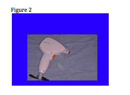
Remember that with most of the lasers that we’re using today, the light piece is “back in the box” and is delivered through some sort of delivery system; however, in the case of some of the mini diode lasers, the light is in the hand piece. (See Figure 2)
Screen Shot 2014-05-26 at 7.50.10 AM

Over the next few years, we are also going to see more and more LEDs. We are going to begin to see more safety features that will be incorporated into the software with touchscreens. For example, you may have a patient who came in for treatment of a wart so the technician turns the dynamic cooling device off. Then, your next patient is coming for treatment of a port wine stain and the technician forgets to turn the DCD back on and the patient gets a blister from the very first pulse. What if there was a tool that provided a warning signal indicating that the DCD was off? Actually that feature is already built into one popular pulsed dye laser.

What about photon recycling? This is a way to capture some of the photons that were wasted. Whenever we use a laser much of the energy is reflected back off the skin surface. By recycling the energy we have a second chance to use those photons. This preserves energy and puts less stress on the system. We will also see more and more scanning technologies as they are becoming increasingly robust. In the future, we will probably see scanners that will find the target and treat it.

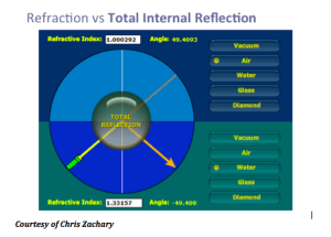
Another concept that Dr Ross discusses is the TRASER (Total Reflection Amplication of Spontaneous Emission of Radiation). The traser is not a laser, nor an IPL. This is one device with many wavelengths that is tunable, has high peak power and variable pulse duration. This device is actually less expensive than a laser and you can change the dye very quickly. The traser uses total internal reflection. We know that if we take light beyond a critical angle, the light will come back towards the same direction.
Screen Shot 2014-05-26 at 8.00.12 AM

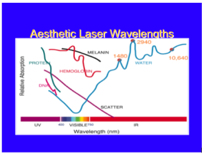
Overall, with lasers in dermatology, you only have to know the chromophore spectra of three targets: blood, melanin, and water. If you know the relative absorption for specific wavelengths for these three targets, you will be a fairly well-armed laser surgeon.
Screen Shot 2014-05-26 at 8.00.00 AM

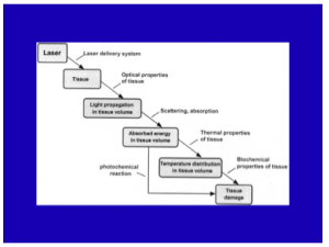
When you model a laser-tissue interaction, you want to look (in your mind) at the way that the light propagates through the skin to the target. You have to get the light to the target. Then, depending on the pulse duration and the wavelength you’re going to have a certain amount of temperature increase leading to a response from that target due to the temperature and time combination.
Screen Shot 2014-05-26 at 7.59.49 AM

What about skin optics? It’s important that you determine the penetration, the absorption and scattering, and the internal dosimetry.

Laser Tissue Interaction Types

There are different types of interactions. The main type that we use in dermatology is thermal, in that we are basically converting light into heat. Other types of interactions include mechanical, chemical, and plasma. Dr Ross feels that one of best ways to learn about laser tissue interaction is through laser hair reduction. The laser comes down, and a certain amount of the light is going to reach the hair bulb, based on the optical properties of the skin. Typically for 1064nm, about 30 percent of the light is going to get about 3mm down which is the typical depth for a hair follicle. Depending on the pulse width, the wavelength, and the fluence, you will have a certain temperature elevation of that hair bulb and some of that heat is going to diffuse to the surrounding skin. So long as the temperature and the time combination is relatively small, you won’t have too much collateral damage—you will only damage the hair follicle.

Selective photothermolysis was a formal termed coined by Dr Rox Anderson 32 years ago. What it says is that if you have the right wavelength, the right pulse duration,  the right target, and sufficient energy,  you will achieve extreme localized heating. This really revolutionized the way in which we treat vascular and pigmented lesions.

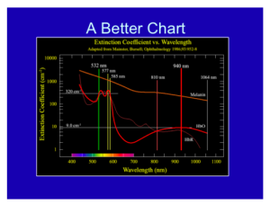
As a dermatologist, you should always remember the graph below, where the wavelength is on the X axis, and you have the relative chromophore absorption in the Y axis. If so, you will be a well-armed laser surgeon. Just like a neurologist looking at an EEG and a cardiologist looking at an EKG, this should be second nature to you.
Screen Shot 2014-05-26 at 7.59.36 AM

How can we exploit laser physics? An example of this is a scar. When we have a scar, there is usually some feature that makes it different from the surrounding skin. You can take a laser and exploit that, whether its redness or pigment in the scar.

This is the temperature equation that basically tells us how hot targets get…the temperature elevation of any target is proportional to the relative absorption of that target for that wavelength of light times the energy div over by a constant. This is very simple concept as its basically energy balanced. Where is the equation??

As you go to longer pulses, the selectivity of the heating becomes poorer but the violent nature of it becomes less.

Fat

Dr Ross states that we are still woefully poor at targeting fat, whether it’s radiofrequency or laser. There are a couple of wavelengths that can be useful for exploiting fat; 1210nm and 1720nm. Those are the wavelengths where fat absorption is in excess of water absorption. However, the ratio of absorption from fat to water is only 1.2 or 1.1 to 1. When we treat a vascular lesion, our absorption rate for the blood is more like 100 to one vs water. So relatively speaking, fat shows poor selectivity, but if you deliver heating and cooling right, there is some selectivity for fat absorption.

Other Key Points

With regards to cooling and heating, we always want to preserve the epidermis and a dynamic cooling device can do so. It is also important to know the photochemistry with regards to chemical reactions and pay attention to these reactions; an example of this can be seen with patients who have previously been on Gold.

Microwaves and Radiofrequency

Microwave and radiofrequency devices are becoming increasingly commonplace within our armamentarium. They rely on heating water, the resistance of water molecules turning causes heat. The  microwave device is around 6GHz. Problems like underarm sweat can be treated with systems like MiraDry. Basically what happens is that the microwaves come down and there is a discontinuity of the dermal-fat junction and it heats up the sweat glands about 4mm below the skin. MiraDry offers another application in clinical practice and can complement other procedures. It also has little downtime.

We are going to begin to see more and more radiofrequency (RF) reactions. It is important to understand SAR, i.e., the specific absorption rate at which energy is absorbed when exposed to an RF electric field. By delivering the right time and the right temperature, one can establish very nice reliable heating of the skin.


MauiDerm News Editor- Judy Seraphine


Tightening Tissue and Zap the Fat

Mathew M. Avram, MD, JD

Dr. Avram provides his clinical pearls regarding the use of lasers in tissue tightening….

The Good News, The Bad News, and Overall Conclusions….

  1. Each of the available technologies can achieve some degree of tissue tightening.
  2. They are the best alternative to surgery.
  3. They are getting better.
  4. For the most part, they are safe in skilled hands.
  5. The benefits, if any, require months to be seen.
  6. Too often, no improvement is seen clinically.
  7. It is difficult to predict who will benefit and who will not.
  8. Tissue tightening can non-invasively provide improvement of skin laxity in a safe manner.
  9. Remember that the results are unpredictable.
  10. The technologies do not approach results of a face lift.
  11. It is very important to educate patients as to the limits of these devices prior to treatment.

Laser Treatment of Pigmented Lesions

Mitchel P. Goldman, MD

Dr. Goldman provides us with some key takeaway points regarding the laser treatment of pigmented lesions…

  1. Nevus of Ota respond to Q-switched laser—we’ve never seen a report of it becoming melanoma; Picosecond is efficacious as well perhaps with fewer treatments.
  2. There are various approaches to treating lentigines—multiple treatments with lasers combined with bleaching agents, sun screen and sun avoidance In addition, resurfacing and chemical peels may also be effective.
  3. Both congenital nevi and aquired nevi can go away with laser treatment; acquired nevi may be easier to remove (sometimes only 1 treatment is needed); Picosecond also shows benefit with perhaps fever treatments.
  4. Melasma—this is one of the most difficult conditions to treat; it typically has a peak in ages 40-50 and is more around the central face. The most important part of treatment is protection from both sun and other light. Laser and IPL treatment is best when combined with skin bleaching agents. We have found Lytera™ to work best.
  5. Infraorbital Pigmentation-Q-Switch Ruby Laser can help with this; PDL and non-ablative and ablative fractionated and confluent resurfacing is also helpful.
  6. What about lentigo maligna? Q-Switch Alexandrite Laser + zyclara may be an appropriate procedure; however, long-term results are unknown; this is also an alternative approach to patients who do not want to undergo surgery.


Fractionated Laser Technologies: Just the Fracts…

Sandy Tsao, MD

In this presentation, Dr Tsao discusses fractional resurfacing and its efficacy in clinical practice. A lot of information has become available to clinicians regarding improving the aging process for their patients.  Fractionated resurfacing technologies have been designed to provide benefits for many aspects of aging by taking the best of the ablative and non-ablative technologies.  In comparison to other non-ablative technologies, fractional non-ablataive resurfacing demonstrates greater efficacy.  It is important to remember that fractional resurfacing is safer for non-facial areas.. It is also safer for darker skin phototypes. , Fractional resurfacing demonstrates a safer side effect profile when compared to traditional ablative devices.. The recovery time is markedly reduced and there are no permanent lines of demarcation to date.

There are a number of fractional technologies available; however, dermatologists should remember that there is not one particular laser that can target all of the concerns regarding photoaging. When you are thinking about these devices, you need to consider what you are actually trying to target. Dr Tsao comments that the ultimate goal in fractional resurfacing  , is to create a localized thermal injury. Thermal injury is thought to be the key for tissue repair. Heat-induced inflammation results in immediate collagen shrinkage and tissue contraction and, subsequently, fibroblast stimulation and neocollagenesis. This concept, Dr Tsao mentions, is very similar to the ablative devices; however, the side effects and down time are much less. There is a zone of irreversible thermal damage and its associated inflammation which must heal before re-epithelialization begins. Prolonged inflammation due to infection, hypersensitivity, or extensive thermal damage due to vigorous treatment can result in complications.

How do fractional resurfacing devices work?

As previously mentioned, there are a number of fractional ablative and non-ablative technologies available,. It is important to match treatment depth and treatment parameters with the clinical  indication(s), i.e., are you targeting pigment (superficial to mid dermis), mild rhytids (mid reticular dermis), moderate rhytids (deep reticular dermis) or surgical scars, burn scars and acne scars (deep remodeling). This is the real learning curve with these devices…you need to know what you are targeting and whether or not you may need a combination of these therapies to achieve your clinical goals.

Each of the devices has a different way of administering the microthermal zones. These devices include continuous motion scanning and traditional lens array stamp pattern, which can be a little bit harder to administer because of the potential of overlap or skip regions.

Non-Ablative  Fractional Resurfacing

Every pulse creates a localized injury or,  microthermal zone (MTZ). You are actually creating columnated areas of heat inflammation. The depth and diameter depend on the type of device that you are using, as well as the parameters selected for treatment. The fractional non-ablative devices create the columnated thermal injury while leaving the normal intact skin in between; therefore, retaining  viable stem cells in between the microthermal zones of injury. This allows for the skin to repopulate much faster, resulting a limited healing period.

Fractional non-ablative laser treatment results in complete re-epithelialization in 24 hours. In human tissue, you can see clear collagen denaturization from papillary dermis into mid reticular dermis. The healing occurs from viable tissue and the zones of spared tissue which contain clusters of epidermal stem cells and Transit Amplifying (TA) cells. What is unique about the non-ablative technologies is that they leave the epidermis intact. The downtime for most of these technologies is about three days of some redness and swelling. This is a very manageable outcome for most patients.

Dr Tsao also comments that over the past two or three years, we have been very fortunate to have a new fractional non-ablataive device available at 1927 nanometers which matches one of the water absorption peaks in the mid infrared spectrum . This wavelength absorption is stronger than most non-ablative wavelengths and weaker than ablative wavelengths. This device fulfills the role to more selectively target lentigines, pigmentation,  actinic keratoses and seborrheic keratoses

Ablative Fractional Resurfacing

These devices work exactly like the non-ablative devices in that they create microthermal zones; however, the big difference is that the epidermis is shed along with a portion of the dermis.  This creates more thermal damage, requiring a longer recovery time. Replacement of the skin surface requires about one week. Patients must be aware of the longer downtime with the fractional ablative devices. Ultimately, what you are aiming to achieve is a healthier more youthful epidermis and dermis.

How does this translate clinically?

If a patient does not want surgery or ablative treatment, then fractional non-ablative or ablative resurfacing could be a viable option. It is important to set realistic expectations with your patients as far as achievable outcomes.  Non-ablative fractional devices will provide improvement of superficial rhytids and dyspigmentation, but not likely provide significant benefit for deeper rhytides.  The fractional non-ablative devices provide marked improvement of atrophic and acne scars.  The fractional ablative devices provide greater benefit for moderate to deep rhytides and dyspigmentation, especially perioral rhytids.

Downtime

It makes a tremendous difference to patients if you can have photos available to show them the expected downtime so that they can make a more informed decision regarding their treatment.

  • Microlaser peel- 7 days of peeling, 2 weeks of pink hue
  • Fractionated non-ablative
    • 1410nm- 1 day mild erythema and edema
    • 1550nm- 2-3 days erythema and edema
    • 1927nm- 4-5 days erythema and edema and pinpoint crusting; 1 week of pink hue
    • 2940nm- 7 days erythema and edema, 2 weeks of pink hue
  • Fractionated ablative- 1-2 days pinpoint bleeding, 7 days erythema and edema; 4-6 weeks of pink hue
    • Ablative-14 days erythema and edema; 2-3 months of pink hue
Clinical Pearls

When treating the eyes, you have to be extremely careful to prevent ocular damage. Less aggressive treatment is necessary  to prevent ectropion formation. It is also important to use  metal protective eyeshields when treating the eyelids. Proparacaine anesthesia and erythromycin ointment to coat the eyeshields can be used for lens placement. It is also important to use particular caution in patients with prior surgical procedure history, such as a prior facelift or blepharoplasty, as the facial anatomy may be altered .

With regards to darker skin phototypes, less is more (fluence, density, passes). You may want to consider a laser test site. You should also stress the need for strict photoprotection and discuss the increased risk for PIH. , You may consider pre-treatment use of retin-a or hydroquinones. You should also use greater cautionwhen using ablative fractional devices, as the risk of side effects is greater.

Remember that when you apply these treatments to non-facial regions you have to be very cautious as there are not as many pilosebaceous units to assist in re-epithelialization, resulting in an increased risk of scar formation when treating these areas with both fractional non-ablative and fractional ablative devices.


Blasting Away Pigment: Targeting Hair, Tattoos, Brown Spots

Michael H. Gold, MD

The tattoo removal market is projected to grow significantly in North America. In 2004, there were 19,900 tattoo removal procedures at an estimated cost of $5.9 million and by 2009 there were 33,900 procedures estimating $14 million demonstrating a 21 percent increase in the market.  There are two major market drivers for the procedure, the first being tattoo popularity. A study at Northwestern University published in the JAAD in 2006 reported that 23 percent of people in the United States have at least one tattoo (69,000,000 people). This was a 30.4 percent increase over the 2003 Harris Interactive study that reported that 16 percent of the US population had at least one tattoo. The second market driver is “buyers remorse” coming from “tattoo regret”-driven by changes in life and/or circumstances. “Tattoo regret” ranges from a reported 15 to an estimated 50 percent of tattoo wearers.

There are multiple, low-cost treatment options for tattoo removal. These include:

  • Cryogenics, i.e., cryotherapy
  • Acids of various kinds
  • Topical retinoids
  • Hyfercation
  • Continuous Wave Lasers
  • Bleaching agents
  • Surgical excision
  • Abrasives with or without chemicals
  • Dermabrasion or salabrasion

However, these methodologies come with many complications such as scars, permanent pigmentation changes, residual tattoo pigment, wound infection, ineffective overall treatment, and can be a painful and slow process.

Laser Tattoo Treatment

There are several advantages to laser tattoo treatment. It is a non-invasive procedure with less pain that produces optimal clearing. With laser treatment, there is a lower risk of scarring and reduced hypo-pigmentation. There is also no change in skin texture, minimal post-operative care and the healing usually takes about one to two weeks. However, there are disadvantages to laser treatment.  Pain is the number one issue (it hurts a lot more to remove a tattoo than to get one), but topical and injected analgesia can be used in conjunction with the laser.  Realistically, it is important to let customers know that as many as 10-15-20 treatments may be required to “satisfactorily” remove a single tattoo (the tattoo will lighten 30-50 percent with each treatment). It is extremely important to set appropriate expectations with you customers and their patients. The treatments can result in hypo-pigmentation, routinely resulting in redness and swelling and punctate bleeding is also a possible (but “normal”) side effect.

As with ANY laser treatment, other more serious side effects are also possible, including–hypo-pigmentation, burns, blisters & scarring.

Pigmented Lesions

The market for pigmented lesion removal is also projected to grow significantly in the coming years. These lesions include epidermal lesions (sun spots, age spots, and melasma) as well as dermal lesions (nevus of Ota/Ito and blue nevi).

Treating Pigmented Lesions with Lasers

Dr Gold comments that both the alexandrite 755 nm lasers and the Nd: YAG 1064 nm lasers are both effective lasers for pigmented lesions. There are several advantages to treating pigmented lesions with lasers, these include the fact that it is a fast procedure; it is a simple treatment technique with minimal discomfort. DCD may or may not be required, depending on the device being used. It usually requires one to four treatments and the lesions darken for five to ten days before they exfoliate. There is improved efficacy with laser treatment because of the thermal effect and the photoacoustical effect.

Several lasers exist for the treatment of both tattoos and pigmented lesions. For pigment, long-pulsed alexandrite lasers are good; Q-Switched lasers are the treatment of choice for tattoos and most pigmented lesions.  It is important to remember that tattoos should NOT be treated with IPLs. Recently picosecond lasers have become available which show, in clinical studies, reduces the treatment time for the overall improvement of tattoos.

R20 Tattoo Removal

R20 involves performing three to four laser treatments in one session.  The result of the R20 method is 50 to 85 percent clearing in a single session without any additional side effects. Laser Tattoo removal treatments form tiny vapor bubbles in the skin (frosting) that looks like fine white powder. Ordinarily, this frost refracts the laser beam preventing it from penetrating the skin. After 15 to 20 minutes, the frost disappears making it possible to do another treatment or “pass”. Dr Gold states that the R20 Tattoo Removal method simply waits for this to happen and lets us treat clients three to four times in one visit. Combining three to four laser tattoo removal treatments during one office visit lets the laser reach deeper than with a traditional single-pass treatment. One R20 treatment would be sufficient to completely remove an amateur black tattoo or for lightening a tattoo for a new one.  For professionally applied black tattoos however, about 50 to 75 percent fading will occur. This is significantly more fading than expected from three to four conventional treatments.  Using the R20 method will cut the total time for removal by half or more.

Conclusions

In conclusion, we know that 25 million people in the US have a tattoo. There are 250,000 women being tattooed each year. Approximately 50 percent of people who have a tattoo look to have them either removed or augmented. The average age of procuring a tattoo is eighteen, often leaving the tattoo as a permanent reminder of their poor decision to get one in the first place. A clinical pearl that Dr Gold tells all of his patients is that it costs more money to remove a tattoo and it hurts more to remove a tattoo then it does to get one.


Combining Lasers with Toxins and Fillers

Suzanne Kilmer, MD

In this presentation, Dr Kilmer reviewed the techniques for combining the various technologies that are currently available for facial rejuvenation in order to obtain optimal cosmetic improvement for patients.

Dr Kilmer stresses the importance of remembering the 5 Rs:

  • Relax
  • Refill
  • Rejuvenate
  • Resurface
  • Reassess
Relax

It is important to relax the skin with a botulinum toxin to keep both the muscles and the skin from moving as much as it otherwise would. If she is going to laser the skin, the results are improved when the skin/muscles are not moving. Dr Kilmer also uses fillers, in conjunction with the toxins and lasers to fill in lines, tighten up the skin and remove brown/red spots.

Clinical Pearl: Never use toxins, lasers, or any other device that can cause significant swelling on the same day. This can result in the toxin migrating to other places where you do not want it.

Refill-Restore Volume Loss

When using dermal fillers, the objective is to restore volume based on a patient’s specific needs. Fillers can be placed in various areas locally such as the nasolabial folds, marionette lines, deep glabellar rhytids, tear troughs, scars, and the nasal bridge. Fillers can also be used globally in the cheeks and temples.  It is important to remember that there may be a lag time resulting in delayed gratification. It is very important to keep in mind that one can’t massage post poly-L-lactic acid (Sculptra) in areas where Botulinum toxin was placed.

Rejuvenation

There are various approaches to rejuvenation. These include:

  • Vascular approach (more specific?)
    • Stimulated by injury to microvasculature, which initiates cascade of events.
  • Pigment approach
    • Targeting melanin can remove pigmented lesions and may also improve textural changes.
  • Thermal approach
    • Non specific heating leads to injury response/wound healing and possible collagen tightening.
  • Fractional approach
    • Nonablative and ablative
Resurface

Dermatologists can resurface the skin with the chemical or mechanical removal of the epidermis. Devices that be used for resurfacing can be both selective and nonselective.

Combination Treatment

Best order

  • Start with toxins to stop movement and relax muscles.
    • Relax frown, smile and lip lines when doing facial rejuvenation
    • Relax DAOs and neck bands when doing fillers, tightening or resurfacing
      • May need less filler and patients are happy sooner with tightening devices
  • Then filler or laser depending on a patient’s specific needs (and ability for downtime)
    • Never do toxins and lasers that cause swelling at the same time because toxins can migrate.
  • Typically end with filler if still needed after toxins and laser
    • Sometimes the combination will diminish the need for filler
    • If able to tell that will need volume, can do before or at same time as laser

Caveats of Combining Treatments

  • Toxin with Filler
    • Can’t massage post poly-L-lactic acid (Sculptra) in areas where Botulinum toxin was placed
  • Toxin with Laser
    • Can’t do toxin same day as Fractional lasers – swelling can lead to migration
    • Can do botulinum toxin with PDL, IPL, CoolTouch, SmoothBeam, Thermage, Titan
  • Filler with Laser
    • Can do filler same day but do first if doing fractional as swelling can mask need for filler.
  • Fractional with RF tightening
    • Same day – do Thermage 1st because need intact skin but when you do the fractional laser the skin may still be sensitive

Combining Fractional with other Devices

  • Fractional laser with other lasers/txs
    • Lentigines – pre-tx QS lasers, KTPs, etc
    • AKs – LN2
    • Sebaceous hyperplasia, nevi – 1450 nm
    • Vascular lesions – PDL, KTP, Alex
    • Downtime from other treatment is simultaneous and shortened
  • Fractional resurfacing with ablative resurfacing
    • Almost always do fully ablative to upper eyelids
      • More tightening/more predictable – do inner canthi
    • Can ablate/sculpt edges of scars, upper lip lines and elevated lesions
  • Ablative fractional and nonablative fractional resurfacing
    • Nonablative fractional to face, ablative to neck for more tightening /crepiness – useful for those with hx ablative resurfacing/chemical peels/dermabrasion

Other combination treatment includes fat loss and tightening, fractionated RF + QS/KTP/PDL and, fractionated US + QS/KTP/PDL.

Reassess

Remember that combination treatment may minimize the need for other treatments and may increase the interval for maintenance. It is extremely important to assess whether or not the patient’s needs have been met. Ask yourself and the patient if new things have become noticeable now that the initial needs have been met? And remember, with time, additional needs may become apparent.

Summary

  • Botox, fillers and lasers can all be used synergistically to minimize the signs of sun damage and aging.
  • Expertise in technique, use of the best possible modalities, and watching for and treating any possible complications will produce the best results.
  • Combining these modalities may obviate the need for more invasive procedures, such as facelift


Fractionated Laser Technologies

Sandy Tsao, MD

There is a lot of information out there and our patients are coming to us with questions about the various devices. Remember, there isn’t one clear-cut way to manage your patients.

What’s the rationale for fractional resurfacing?
  • Safer for non-facial areas; beard area
  • Safer for darker skin phonotypes
  • Efficacy greater compared with other non-ablative technologies
  • Safer side effect profile c/w ablative devices
  • Recovery time markedly reduced
  • No permanent lines of demarcation to date
  • Non-ablative and ablative devices

There are a variety of fractional technologies for rejuvenation currently available to the practicing dermatologist.

The Goal of Thermal Injury

Thermal injury is thought to be key for tissue repair. Heat-induced inflammation results in immediate collagen shrinkage and tissue contraction and subsequent fibroblast stimulation.  There is a zone of irreversible thermal damage and associated inflammation must heal before re-epithelialization begins. Prolonged inflammation due to infection, hypersensitivity, or extensive thermal damage due to vigorous treatment can result in complications.

How does fractional resurfacing work?
  • One pulse produces one MTZ
  • Thermally induced coagulation, not vaporization
  • Columnar shape
  • Diameters are variable
  • 1:5 width to depth ratio
  • 5-48% total skin coverage – each treatment

It is important to remember to match treatment depth with the indication.

Pigment-Superficial to mid dermis

Mild rhytids- Mid reticular dermis

Moderate rhytids – Deep reticular dermis

Surgical scars and acne scars- Deep remodeling

 Fractional non-ablative laser treatment in a cross section of human tissue demonstrated complete re-epithelialization within 24 hours. There was clear collagen denaturation from the papillary dermis into the mid reticular dermis. Healing occurs from viable tissues. Zones of spared tissue contain clusters of epidermal stem cells and Transit Amplifying (TA) cells.

Ablative Fractional Resurfacing

These work like the nonablative devices; however, you lose the epidermis.

  • Ablative tissue coagulation with loss of stratum corneum
  • Multiple microthermal zones of injury surrounded by viable tissue
  • Resurfacing with replacement of skin in 5-7 days
  • More thermal damage with longer healing time
  • Immediate and delayed therapeutic benefits greater
    • Epidermal and dermal coagulation for resurfacing
    • Collagen denaturation for tissue remodeling


The downside to laser treatment can be the downtime associated with the procedures and patients should be aware of this. Estimated downtimes are:

Microlaser peel– 7 days peeling, 2 weeks of a pink hue

Fractionated non-ablative

1410nm- 1 day of mild erythema and edema

1550nm- 2-3 days of erythema and edema

1927nm- 4-5 days of erythema and edema

2940nm- 7 days of erythema and edema. 2 weeks of pink hue

Fractionated ablative– 7 days of erythema and edema; 4-6 weeks of pink hue

Ablative– 14 days of erythema and edema; 2-3 months of pink hue

Laser Treatment of the Periorbital Region

When treating  around the eyes with laser therapy, less aggressive treatment is needed. When treating the lids, a metal protective lens is necessary. For lens placement, use alcaine anesthesia and erythromycin. Be sure to use particular caution in patients with prior surgical history.

Darker Skin Types
  • Less is more (Fluence, density, passes)
  • Consider a laser test site
  • Stress the need for strict photoprotection
  • Discuss the increased risk of PIH
  • Consider pre-treatment use of retin-a or hydroquinones
  • Use caution with ablative fractional devices

Charities and Laser Treatment

Michael Gold, MD

There are many charities throughout the world that are providing laser treatment for scars due to injuries and/or accidents to those who cannot afford the treatment.

The Wounded Warrior Project, here in the United States, is providing laser treatment to our wounded troops. This effort is really helping to improve the quality of life of veterans.