Advances in Treating Varicose Veins and Telangiectatic Leg Veins

Mitchel P. Goldman, MD

Dr Goldman is a Volunteer Clinical Professor of Dermatology at the University of California in San Diego, Immediate Past-President of the American Society for Dermatologic Surgery (ASDS), and an expert in Sclerotherapy publishing 5 textbooks and dozens of medical papers on varicose and telangiectatic leg veins. In this presentation, he provides us with an update on the treatment of varicose and telangiectatic leg veins.

Thirty percent of the population will have varicose and telangiectatic leg veins by age twenty. As we age, the incidence increases, i.e., 80 percent of the population will have varicose and telangiectatic leg veins by age 80. There are many reasons for developing varicose veins. The incidence among men and women is about equal. It is important to remember that up to fifty percent of varicose vein patients will develop DVT, SVT, and/or ulceration in their lifetime.

Dr Goldman feels that the more factors you have, the more likely you are to get varicose veins.

Screen Shot 2014-12-26 at 7.47.31 AM

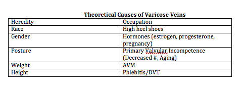
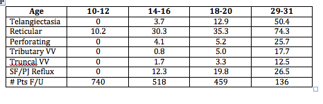
When you look at longitudinal studies of large populations (Bochum Study—Schultz-Ehrenberg, 2004), you will see that the veins start out as spider veins and then gradually, the older you get the more reticular or truncal varicose veins appear.

Screen Shot 2014-12-26 at 7.47.20 AM

Why do people come to us for their varicose veins? They come primarily not because of symptomatology, but for cosmetic reasons. Most physicians think that veins are just a cosmetic procedure and not a real medical problem, but this is an actual medical condition. In Dr Goldman’s office, even when leg pain is not a presenting concern, it is not uncommon for patients to tell him that their legs feel better after treatment.

Varicose and Telangiectatic Leg Veins: Symptoms

  • Heaviness
  • Aches and Pains
  • Night cramps/Restless legs
  • Ankle edema

Why is there pain? There’s pain because the venous hypertension, i.e., veins that you can see on the surface, are actually surrounding and pressing on nerves. Venous reflux can cause varicose veins, skin changes, skin ulcers and swollen legs. This is why dermatology is the specialty that should be treating this condition. In Europe, it’s the dermatologist who is the phlebologist.

Current treatment options include:

  • Compression stockings
  • Sclerotherapy
  • Phlebectomy
  • Stripping and ligation
  • Endovenous laser
  • Endovenous radiofrequency

When treating patients with varicose veins, insurance companies will first ask you if the patient has failed compression stockings—implying that the patients should be wearing these stockings for the rest of their life. If patients are complaining of pain, an easy way to see if it’s caused by the veins is to put your patients in graduated compression stockings. If the pain goes away, then you know that sclerotherapy or treatment of the veins is going to make the pain go away as well.

A study by Schul et al compared compression versus sclerotherapy for patients with isolated refluxing reticular veins and telangiectasia. They looked at 50 women with symptomatic veins with normal saphenous and deep veins. Ninety percent of the women required OTC analgesics. The subjects were randomized to thigh high 20 to 30 mmHg graduated compression for six weeks versus sclerotherapy. All compression patients crossed over to sclerotherapy. The researchers found that graduated compression improved aching, pain, cramps, and restlessness and sclerotherapy improved everything plus made the veins go away. (Scul et al. Phlebology. 2011;26:148.)

When you treat varicose or telangiectatic leg veins, you have to be logical and do it in an order. You can’t just hit or miss with vein injections and expect a good result. It would be the same analogy as if a patient had a skin cancer on the face—you would not just treat little bits here and there.

A Logical Progression

  • Begin with cut-off of reflux from SFJ/SPJ
    • Endoluminal RF or LASER Closure or
    • Foam Sclerotherapy
  • Ambulatory phlebectomy or Foam Sclerotherapy of veins > 4mm
  • Sclerotherapy of remaining veins
  • Then consider LASER or IPL

When a patient presents with telangiectasias in one little area, you need to remember that telangiectasias are fed by deeper reticular veins all of the way into the deep venous system. Everything is connected. When Dr Goldman treats a patient, he doesn’t do a section at a time, nor does he bill by the amount of solution that he’s using. He treats per leg. It is imperative that you treat the entire venous system in one leg in order to minimize the number of treatments.

Screen Shot 2014-12-26 at 7.48.10 AM

Screen Shot 2014-12-26 at 7.48.33 AM

What gets us into trouble when we don’t know the anatomy is that we have to figure out where the veins are coming from. There are times when the veins don’t come from the incompetent great saphenous vein. Dr Goldman uses a duplex machine to determine where the veins are coming from.

As Dermatologists, we tend to see the lateral subdermal plexus. When patients present with little telangiectasias on the lateral aspect of the thigh, which is very common, we must realize that it’s all coming from an incompetent reticular vein. You must treat the entire leg.

If you leave a vessel open after it’s been injured, your body is going to reconstitute it and the endothelial cells are going to keep trying to reestablish the vessel. If you compress the vessel after you’ve injured it, you can decrease the extent of thrombosis, which decreases the risk of recanalization and pigmentation allowing for the vessels to go away quickly. Walking is another way to do compression. When you walk, you’re basically siphoning off the blood flow from the superficial venous system into the deep venous system and that’s doing a physiologic form of compression. You want to minimize the thrombosis as best as you can.

Screen Shot 2014-12-26 at 7.48.44 AM

You want to have patients who are going to follow your directions as compression really does improve outcomes. Dr Goldman only uses thigh-high stockings for the patients. He stocks them in his office because the pharmacies and medical supply offices do not always know how to best measure for stockings correctly. The stockings must be applied immediately after treatment.

What is sclerotherapy?

Sclerotherapy is a controlled thrombophlebitic reaction that began hundreds of years ago. There are two solutions approved by the FDA that can be used—sodium tretadecyl sulfate (STS) and polidocanol (POL). Glycerin is also an approved solution.

Sclerosing Solution Matched to Vessel Size

  • Telangiectasias < 1mm in diameter
    • Very small volumes of hypertonic saline and/or dextrose
    • 0.1% – 0.25% STS liquid
    • 0.25% – 0.5% POL liquid
    • 72% Glycerin mixed 2:1 with 1% lidocaine +/- epi
  • Reticular veins > 1mm in diameter
    • 0.25% – 0.5% STS foam
    • 0.5% – 1.0% Pol foam

When you start injecting a vein, you have to go back to your high school physics. You have to fully inject until you stop seeing solution in the vein because once you stop seeing where the solution is flowing, it’s flowing deep—where you don’t particularly want it.


  • Volume of a vein-Vv=ttD2/4L (D= internal diameter and L = length)
  • 2ml into a 5mm travels 10cm; 0.5ml into a 0.2mm vein travels 6cm

Detergent solutions (sodium tetradecyl sulfate and polidocanol) are very efficient. They strip off the proteins from the endothelial cell causing its destruction.

Screen Shot 2014-12-26 at 7.48.54 AM

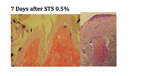
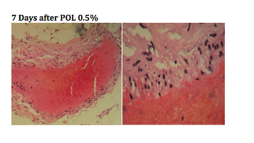
You can see, from the biopsies above, that there is a total wipe out of the endothelial cells. Using the same dosage of polidocanol, we don’t see the wipe out of the telangiectasia, it basically just attenuated the endothelium.

Screen Shot 2014-12-26 at 7.49.05 AM

When you’re using STS versus POL, the concentration is essentially two to one. POL is one half of the potency of STS. Both solutions are very comparable in efficacy.

Parsi and colleagues studied the in vitro effects of detergent sclerosants on antithrombotic mechanisms. They found that STS is antithrombolytic and does not destroy plasminogen. Reinjection of STS into a sclerosed vein can accelerate recannalization. POL has no effect on coagulation factors. If the sclerosant does not destroy the endothelium, it will stimulate inflammation, which produces t-PA with fibrinolysis. Parsi, et al. Eur J Endovas Surg. 2009)

Dr Goldman does NOT use hypertonic solutions for a few reasons. Osmotic agents like HS hurt and they only act within their osmotic gradient leading to a much higher risk of recurrence as well as pigmentation and pain.

Glycerin, in and of itself, is a hypertonic solution, but it is very safe. Dr Goldman has never seen pigmentation or ulceration from it. It is available and can be compounded from a USP pharmaceutical grade glycerin.

Screen Shot 2014-12-26 at 7.49.18 AM

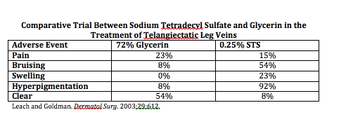
Glycerin has comparable efficacy with less adverse effects. Dr Goldman usually adds lidocaine to the glycerin; the lidocaine does not affect the efficacy of the glycerin; however, it does minimize the pain.

Compounding Solutions

Compounding solutions are not to be done in sclerotherapy. This can open up an amazing amount of lawsuits that will not be defended by your malpractice.   Why does Dr Goldman use compounded glycerin? It has proven to be very safe and is basically the same product that neurosurgeons are using in the brain for cerebral edema.

Foaming

There are advantages to foamed sclerosants. They are increased in volume but the total sclerosant injected is decreased. The highest concentration contacts vessel walls causing total obliteration of the vessel. There is a very slow washout and long contact with the intima. The only problem with foaming is that one third of the population has patent foramen ovale causing the foam to go into the arterial system. It is very safe to foam and it is safe to have nitrogen bubbles in your body. Dr Goldman has been using foam for twenty years and has only had two patients complain of a headache or slight loss of vision for just a few seconds.

Conclusions

You really have to look at your patients and evaluate the results. Taking photographs is very important. It is important to have good communication with your patients in order to set realistic expectations.

Patient Preparation

  • Eat before hand to avoid vasovagal response
  • Don’t shave legs or use moisturizers day of treatment
  • Wear shorts during treatment sessions
  • Bring pre-fitted GCS to visit and FU
  • Patient is recumbent, ideally on power table

Equipment and Supplies

  • Good light   vein light   polarizing light
  • Comfortable table
  • Comfortable chair for you
  • Digital Camera
  • Sclerosing agents
  • Normal Saline
  • Alcohol soaked cotton balls
  • 4×4 Gauze sponges
  • Graduated compression Stockings
  • Needles
  • Syringes
  • (3 and 5ml + 2 way connector)
  • Mayo Stand
  • Hazardous waste container
  • Gloves
  • Chucks

In thirty years, Dr Goldman has never had major problem; however, allergic reactions and anaphylaxis can occur, so you must be prepared with resuscitation equipment. You should have an office protocol in order to deal with emergency situations.

Injection Technique for Telangiectasias

  • Use 30 gauge needles ½ inch long
  • It usually takes 0.1-0.2 cc’s to inject a spider vein
  • Avoid forceful pressure
  • May bend needle – 30 degrees
    • 30 gauge, plastic hub disposable
    • 33 gauge dull quickly, bend too easily, not disposable
  • Can’t draw blood back, and not necessary
  • Inject very slowly, with little pressure, until blood cleared
  • Graduated compression hose (30-40 mm Hg) for 1 week
    • Reduces pigmentation
    • Better response
  • Identify patterns and groups, e.g. lateral venous plexus
    • Use additional tricks if necessary
  • Begin injections at logical starting points (reflux sites) or base of telangiectatic web
  • Keep your eye on injection site at all times
  • Minimize volume of solution per site
  • Recognize earliest bleb formation and stop!
  • Use minimal sclerosant concentration

Again, the most important thing is that you do this logically. You want to eliminate the high-pressure reflux first. Treat the small varicose and reticular veins initially—associated telangiectasias should be treated on the same day. You want to treat from proximal to distal and treat the entire length of the vein in one session. Dr Goldman follows up with the patient six months post-procedure for maintenance.


The Aesthetic Consult

Kent Remington, MD

Dr Remington is an internationally recognized leader in the aesthetic dermatology industry. In this presentation, he discusses his novel approach to the aesthetic patient. Dr Remington’s aesthetic consult is quite different from that of other dermatologists. We all want to reinvent ourselves and it’s necessary to keep up with the ever-evolving field of aesthetic dermatology.

How can we get patients who are on a budget to go ahead and agree to a fairly comprehensive procedure/project? It’s all in the consult. It is important, as physicians, to recognize that patients are skeptical, suspicious, leery, tentative, intimidated, apprehensive, tense, cautious, and so on when it comes to facial rejuvenation. Dr Remington stresses the importance of “peeling all of these layers off” to be able to have some connection and engage with your patient(s). Remember “one size fits none.” Patient’s are not paying for cost, they’re paying for good results. If you look at patient’s animations, not everyone is the same. We have to be able to teach our patients what their face really needs. Dr Remington feels that most injectors just follow what everyone else is doing. It’s easy to follow what everyone else is doing; we are extremely busy in our clinical practices and we can tend to confuse being busy with accomplishment. The old aesthetic rule was “if it ain’t broke, don’t fix it.” The new rule is “if it ain’t broke, break it and make it better.”

The aesthetic consult requires that you look at the “whole face.” Attention to detail should be your major focus. Patients tend to have selective focus. They come into your office with a focus on their nasolabial fold or their frown zone and that’s it, they don’t see anything else. Patient’s perception of reality and reality are often quite different…we look with our eyes and see with our brains.

Patients expect value for their dollar and they expect to be treated well, i.e., they don’t want good results, they expect outstanding results. This requires careful planning on the part of the physician by balancing neuromodulators, fillers, surgery, lasers, energy devices and fat busters with cost. Its not about dollars, its about results. Patients are willing to pay for good results. Dr Remington has five nurses who work with them and they take a very detailed approach to facial reflation and contouring.

Faces are all about genetics, genetics are about biology, biology is about physiology, physiology is about physics, and physics are about mathematics; therefore, faces are all about math. You have to have a plan in mind, eg., where is the peak of the brow? What is the width of the lip? As far back as 1934, makeup artists were given guides to the face on where/how to best apply makeup based upon facial dimensions. Dr Remington refers to his process as “facial beautiPHIcation.” “Creating great results is a result of good mathematics, I am not talking about left brain math, but right brain math”

You want to uncover the “real reason” that your patient is interested in looking better. You can discover the reasons not by questioning, but by intuitively interacting with your patients.

Dr Remington asks every patient to bring in a good photo of him or her. Usually, this is a picture that was taken in his/her twenties. Dr Remington isn’t trying to make them look twenty, but he wants evaluate past symmetry,balance, proportion and balance or lack thereof. A picture makes it much easier to create what you’re trying to show them. Often times, he will split the face in a computer screen set up to show the patients youthful photo beside the current one. This helps patients see clearly why we need to look at and treat the whole face in a planned restoration project. Not to make them look different, but to restore what they used to look like A dedicated camera room with a high-resolution monitor is a MUST, according to Dr Remington because this is where he can do much of his education and visual perception for his patients. Sometimes Dr Remington will convert the color photos to black and white because it is easier to show contrast, volume loss and wrinkles.

Good results come from commitment to a detailed consult,.In North America, many people come from a combination of ethnic backgrounds. Dr Remington spends a lot of time with his patients discussing their genetics. Why is this important? We want to find out the positive part(s) of their genetics. He tries to not only retainthat part but also, accentuate it. Aesthetic physicians need to train their eyes to be like video cameras with Panavision friendly focus; but the aim is on information and figuring out everything about the patient. You must understand where the patient is coming from—this comes from connecting and engaging with your patient.

Dr Remington and a colleague found that there are seven types of consulting styles.

  • Socializer
    • Initially impress patients
    • Rarely get past the social part
    • A lot of odour, little substance
  • Consultants
    • Good listeners
    • Go with patient’s monotherapy focus
    • Succumb to patient’s unreasonable requests
  • Closers
    • Smooth, slick style
    • Up sell rather than up serve
    • A lot of dancing to seal the deal
  • Storytellers
    • Love to detail patient case studies
    • Forget to focus on the bigger picture
    • Try to endear themselves to the patient
  • Focusers
    • Know the science of all their products and devices
    • Discuss and treat selective focus areas of the face
    • Tunnel vision
  • Narrators
    • Love to listen to themselves talk
    • Discuss procedures like robots
    • Do not recognize that “one size fits none”
  • Face whisperers
    • Engage the patient and ease their apprehension
    • Up serve rather than up sell
    • Calm, focused assertive energy

Patients can sense your uncertainty. You have to present with confidence so that your patient(s) understand and agree with the procedures that you recommend. Remember that words are extremely powerful. “Words are the most powerful drug used by mankind. Not only do words infect, egotize, narcotize and paralyze—they get into color even the minutest of brain cells.” (Rudyard Kipling) Words are hypnotic and they can make a huge difference to your patient.

Dr Remington has discovered that an aesthetic patient is driven to look more youthful not on the size of their wallet or their hormone level it’s in their DNA. You have to determine which patients are aesthetically wired, and which are not.

Men and women focus on different things when they look in the mirror. Women tend to focus on their eyes and their lips and men tend to focus on spots. Again, patients have selective focus. What they see and what is actually real are often quite different. As aesthetic physicians, we have to gently teach patients what we see. We are image enhancers. We want to find and point out the patient’s positive facial features. This is important for our patient’s self image, self-esteem and self-concept. How we think we look has a direct influence on our appearance.

It is important to take a “global approach” to facial restoration and beautiPHIcation. Physiognomy (face reading), personology (reading personalities) and phenomenology are becoming more and more a part of our interest as aesthetic physicians as this gives us the information we need to know about our patients.

Clinical Photography

You have one opportunity to get a baseline, pre-treatment photo. As previously mentioned, a dedicated camera room with a high-resolution monitor is highly recommended. You want to evaluate your patients in good lighting. Start early with your photography, you can teach your patients from the photos.

One of the most important things that Dr Remington does for teaching is insisting on the patient bringing in his/her old picture so he can split the face to show the difference over the years.

If you look at aesthetic clues about who is interested in aesthetics, there are verbal clues, non-verbal clues, and visual clues—be observant. You need to recognize patients who have had procedures in the past e.g., prosthetic dental work, face lift, rhinoplasty, those who style/dye their hair, patients with special events or milestones coming up such as a wedding or anniversary, and patients with special circumstances, e.g, their partner is younger or a milestone birthday.

In conclusion, it is imperative that you to take a global approach to your patients. A combination of procedures may be necessary for optimal results. Remember to connect with your patients and engage them in the process as our current aesthetic patients world wide expect outstanding clinical results.

MauiDerm News Editor-Judy Seraphine

Applying Laboratory Breakthroughs to Treat Pediatric Skin Diseases: Part 2

Amy Paller, MD, MS

Biologics as Targeted Therapy

Genetic advances are teaching us more about psoriasis as a disease state. The discovery of pathways that influence psoriasis have led to treatments such as TNF alpha inhibitors, interleukin-23 inhibitors, and IL-17 and its receptors. We have also learned that CARD14 mutations are a cause of familial psoriasis/PRP. One of the ways that CARD14 works is through the activation of IL-23 signaling and Th-17 expression, leading to the use of ustekinumab, a commercially available agent that targets IL-12/-23 with success.

Through exome sequencing, we have learned more about generalized pustular psoriasis. There is a deficiency of the interleukin-36 receptor antagonist, DITRA, in hereditary pustular psoriasis (and some cases of “sporadic” generalized pustular psoriasis show mutations in the IL-36R antagonist). This discovery suggests that activated IL-36 (or its related compound, IL-1) may be an important target in pustular psoriasis.

What about alopecia areata? We have learned from some excellent genome-wide association studies (GWAS) that there are several loci that are increased in this disease, one is which is CTLA4. There are ongoing trials of abatacept (CTLA4-Ig initiated) and rixolitinib (JAK 1/2 inhibitor to suppress IL-15 activity), but an early report of JAK1/2 inhibition showed great promise.

Targeting Functional Pathways

We can use systemic administration of small molecule inhibitors to suppress signaling activation. This can be seen with the use of vismodegib in basal cell carcinoma and basal cell carcinoma syndrome (BCNS) (activation of hedgehog signaling) and oral rapamycin in tuberous sclerosis (activation of mTOR signaling). GNAQ mutations cause the majority of blue nevi and 90 percent of cases of Sturge-Weber syndrome and nonsyndromatic portwine stains. GNAQ mutations have now been shown to activate PKC and MAPK but not AKT or mTOR. This observation suggests that an inhibitor of PKC or MAPK signaling might be effective if applied topically. With epidermal nevi, activating mutations have been detected in the genes encoding RAS family members or affecting FGFR3/PI3K/AKT signaling. Differentiating the activated pathway could influence decision-making about the most appropriate inhibitor to apply topically in the future. G13R and Q61R or Q61L Hras mutations in melanocytes cause Spitz nevi and speckled lentiginous nevi, yet another situation in which a targeted inhibitor which blocks Ras signaling might be considered as therapy.

Protein Replacement Therapy

Intradermal or intravenous introduction of recombinant collagen VII into RDEB (recessive dystrophic epidermolysis bullosa) mice leads to the deposition of collagen VII at wound BMZ and decreased skin fragility. Recently, FDA approved a trial of injected collagen VII in patients with RDEB. Future investigations might involve topically applied recombinant collagen VII to wounds or even intravenous delivery to reach mucosal sites as well. Interestingly, studies in mice without epidermolysis bullosa suggest that even normal wounds heal more readily if collagen VII is administered, broadening the potential value of topically delivered collagen VII.

Replacement; however, may not be sufficient. In CHILD syndrome, the cholesterol biosynthetic pathway is blocked, leading to deficiency of cholesterol but also accumulation of toxic metabolites. Application of topical cholesterol to skin is not sufficient, but use of a statin to block both the accumulation of toxic metabolites, as well as replacement of cholesterol leads to dramatic improvement in affected skin using this “pathogenesis-based” therapy.

Summary

New technology has facilitated the advancement in our knowledge about gene function and the effects of gene alteration, including for some of the mosaic disorders. These discoveries have translated into new therapies for patients with genetic disorders and will help with innovative treatment in the future, whether personalized gene-based or pharmacologic therapy. While there are new discoveries to be made, one of the exciting new frontiers involves epigenetics—unraveling how and why genes are turned off with cell and tissue specificity – and then translation this information towards treating human disease.

Infectious Diseases: Update on the Management of Onychomycosis

Aditya K. Gupta, MD

Dr Gupta, an expert in the management of nail disorders, discusses clinical advances with regards to the management of onychomycosis.

Factors Affecting Diagnosis

It is very important that we correctly diagnose onychomycosis. For the first time in over twenty years, we are able to use techniques other than light microscopy and culture. Molecular biology can be used to accurately diagnose multiple organisms that may cause onychomycosis. Dr Gupta has learned, through his lab, that often we may have mixed infections of two dermatophytes, a dermatophyte and a non-dermatophyte, or two non-dermatophytes. PCR analysis is more sensitive than culture, so it has a greater ability to identify mixed infections.

Screen Shot 2014-12-12 at 5.22.53 AM

Treatment

Oral Therapies

Onychomycosis can be managed with topical therapy, oral therapy, and/or devices, i.e., lasers. In terms of oral therapy, terbinafine is the gold standard. Itraconazole is also indicated for the treatment of onychomycosis and fluconazole can be used off-label.

There are two new oral drugs in development—posaconazole and albaconazole.

Screen Shot 2014-12-12 at 5.22.41 AM

It is important to recognize that onychomycosis is a difficult disease to treat, so you should consider the fact that you may need booster therapy or extra treatment around six to nine months from the start of therapy. For example, with terbinafine, giving 250mg per day for four weeks and repeating that at nine months and twelve months.

Topical Therapies

Efinaconazole, a triazole, which was just approved in Canada, has a low MIC for dermatophytes, yeasts and non-dermatophyte molds and relatively low keratin binding to facilitate transungual penetration. With regards to mycological cure and complete cure, data for efinaconazole demonstrated 54 percent and 17 percent cure rates, respectively versus 32 percent and seven percent, respectively with ciclopirox.

Tavaborole, a broad-spectrum benzoxaborole antifungal, has a low molecular weight and low lipophilicity facilitating penetration into the nail plate. At week 52, tavaborole demonstrated a 34 percent mycological cure and an eight percent complete cure as compared to 32 percent and seven percent, respectively with ciclopirox.

We need to recognize that there are differences in the severity and population of participants between the studies looking at the efficacy of efinaconazole and tavaborole; therefore, precluding a head-to-head comparison.

Differences Between Efinaconazole and Tavaborole in Pivotal Phase III Studies

  • No upper age limit for tavaborole
  • Nail trim: tavaborole allowed no more than 1mm nail beyond hyponychium
  • Japan not included in tavaborole study
  • Efficacy of vehicle for efinaconazole 2x that of tavaborole

Luliconazole is a topical imidazole approved in the United States for the treatment of tinea pedis. There are phase II/III trials currently underway studying its efficacy for onychomycosis.

Device Therapy—Lasers

There is insufficient data to determine if lasers are fungicidal in onychomycosis, especially as the precise mechanism of action remains unclear. The most likely effect is a selective photothermal effect, although we cannot rule out a photoacoustic, photochemical, or photomechanical effect. Keep in mind that the fungus has to be heated to at least fifty degrees centigrade for at least ten to fifteen minutes or 60 degrees (or even 70 degrees) for shorter periods of time. The aim of selective photothermolysis is the create very high increases in temperature in the fungi while the water content in the dermis and the blood vessels surrounding the nail bed allows for the dissipation of heat to prevent pain and tissue necrosis. The pulse format of the laser is critical for ensuring that heat remains confined and elevated in the fungi, while providing sufficient time for the surrounding tissue to dissipate the heat,

In the United States, the division of the FDA that approves lasers is different from the division that approves anti-fungal treatments. Lasers are indicated for use for the temporary increase of clear nail in patients with onychomycosis—this is quite a different approval as it is a cosmetic approval that does not look for mycological or complete cure. These lasers have not been subject to the same stringent standards that we expect of topical and oral therapies. There have been a number of open, single assignment clinical trials conducted using laser systems; however, randomized, controlled studies are needed to determine if laser is an effective treatment

Prevention of Recurrence and Reinfection

Recurrence rates can be anywhere from 11 to 35 percent or even higher. Remember that fungi can be transmitted to uncontaminated clothing in the wash. It is important to remind patients to wash their clothes and shoes in hot water (greater than 60 degrees) for more than 45 minutes. Ozone can be used to sanitize shoes and sports equipment of dermatophytes, yeasts, and non-dermatophyte molds. Ultraviolet light can also be used to sanitize shoes and reduce fungal burden.

Factors Contributing to Recurrence and Reinfection

The fungus that causes onychomycosis is not just dermatophytes or non-dermatophytes, but mixed infections as well. We have also shown that T. rubrum is not just T. rubum; there are minute-based pair differences that produce different strain types. We have found that strain type switching may contribute to the failure of oral terbinafine.

In summary, terbinafine remains the gold standard for treating onychomycosis. Both continuous and pulsed regimens are beneficial with an optimal pulse regimen of four weeks on, four weeks off and four weeks on. We have found that using this strategy, taking into account the pharmacokinetics of the drug, we can achieve fairly high cure rates. Itraconazole 200mg melt extrusion tablet may lead to better compliance rates versus two 100mg capsules. Don’t forget about booster therapy!

Fluconazole, while off-label in the United States, can be used at 150mg/week for longer than six weeks is also effective. The efficacy for posanoazole and albaconazole is similar, but not superior, to that of terbinafine.

We also have new topicals for onychomycosis—efinaconazole and tavaborole. It remains to see whether or not they will be used as monotherapy or in combination. Perhaps they could be effectively used in patients who fail oral terbinafine. They may also be used to prevent early recurrence of onychomycosis. There are a lot of potentials for the topical therapy.

As dermatologists, we use lasers for many things. We still don’t know enough about the use of lasers for the therapeutic cure of onychomycosis.

Applying Laboratory Breakthroughs to Treat Pediatric Skin Diseases: Part 1

Amy Paller, MD, MS

At Maui Derm 2014, Dr Paller discussed how laboratory breakthroughs have enabled us to better treat skin disorders in the pediatric population. These discoveries come from the understanding of disease pathogenesis and our seeking of targeted interventions.

New discoveries and new technologies, e.g., gene sequencing, and analysis of RNA and protein expression, are increasing our understanding of skin disease and aiding in the development of new therapies.

Unraveling the Cause of Monogenetic Disorders

Discoveries from Sanger sequencing revolutionized prenatal diagnoses such as amniocentesis, chorionic villus sampling, and more recently for a limited set of disorders, maternal blood sampling. Preimplantation diagnosis is a technique that utilizes in vitro fertilization to pull out a single egg, extract the DNA, and distinguish whether or not the fertilized egg is normal or not and, thus, implant the eggs that will be normal.

Most dermatologists are familiar with Deep-Sequencing or “next-generation” sequencing technology, which is transforming our understanding of genetic diseases and how we can treat them.

Can next-generation sequencing help patients?

By simultaneously analyzing all candidate genes, we can detect low frequency mutations in known genes, mutations in newly associated genes, and low frequency SNPs in thousands of samples. We can also find genotype-phenotype correlations based on alterations in multiple genes. We can analyze epigenetic changes like methylation at base-pair level, histone modifications and protein-DNA interactions. This may provide us with a greater ability to predict responses to medications and personalized gene therapy.

Technologies in Trials Today

Gene therapy: Ex-vivo cell therapy

Researchers at Stanford are using this technology for recessive dystrophic epidermolysis bullosa (RDEB) in adults. One potential risk of ex vivo gene therapy is that the introduced protein may be viewed by the immune system as foreign, leading to a problem with the creation of autoantibodies. At Stanford, the researchers are only accepting patients (~60% of individuals with RDEB) who have the N-terminal domain of collagen VII—the immunogenic domain. This decreases the chance of the patient developing an autoimmune disorder. In these studies the concern about insertional oncogenesis (ie, that the viral insertion, which is random, could lead to interruption of an important tumor suppressor gene) is mitigated by the fact that any tumor that might developed would be highly visible and easily managed. On question is whether spraying the corrected keratinocytes onto a wound bed could be a substitute for grafting skin; this techniques has been used to introduce normal keratinocytes into graft sites for burn patients (Gerlach et al. Burns 2011;37:e19).

Stem Cell Therapy

Several years ago, the University of Minnesota began pioneering bone marrow transplants on children with RDEB, leading to evidence of incorporation of autologous stem cells, deposition of collagen VII at the dermal-epidermal junction, and some improved healing. Subsequent studies have used newer techniques to decrease the risk to patients and increase efficiency, including extending the therapy to junctional EB (JEB) patients. It is important to note that this procedure still carries a risk with variable success and slow improvement.

Revertant Mosaicism: “Natural” Gene Therapy

This term refers to a spontaneous correction of a loss-of-function mutation, leading to “carrier” phenotype. This has been seen extensively in patients with JEB because of mutations in collagen XVII, but has now been described in several other genetic disorders of the skin. This “natural gene therapy” may lead to expanding a small biopsy of patient cells to introduce as a graft or through use of iPS cells (see below) without requiring immunosuppression because the cells are otherwise the patient’s own cells.

Induced Pluripotent Stem Cells (iPS)

iPS cell technology allows one to take any cell (e.g., keratinocytes, fibroblasts) and de-differentiate it into a stem cell using well-known factors. You can then use other factors that will differentiate it into any type of cell that you want. As noted above, with regards to revertant mosaic areas, you can take those cells directly and turn them into stem cells and give someone back his own cells.

Many genetic diseases are dominant negative disorders, i.e., a genetic change in one allele is enough to disrupt function. Small interfering RNA (siRNA) is an approach to suppress the express of a target gene and has already been described in a human trial as a viable means to suppress gene expression and lead to clearance in pachyonychia congenita (Leachman et al. Mol Ther. 2010;18:442). siRNA specifically directed against the KRT6a mutation in a patient was injected into a tiny area of the plantar keratoderma using a left-right, double-blind analysis. Despite clear improvement, the injections were very painful, emphasizing the need for improved techniques for the delivery of genetic material through the epidermal barrier. Protrusion array devices utilize microneedles, often dissolvable, as a painless way to deliver genetic material or protein through the barrier. New creams that can be applied to the skin, including with the use of nanotechnology, are also under development as a means to deliver siRNA. TALENs and CRISPrs induce site-specific, double-stranded DNA breaks, then allow homologous-directed repair with the normal sequence. Although early in development, these techniques hold the promise of delivery of the normal sequence without the risk of random insertional oncogenesis.

Proteomic arrays (amino acid sequences encoded by mass spectrometry) and phosphoarrays (screens for activated proteins.) allow us to look at proteins directly and are great for drug discovery. ELISA assays have also become more sensitive. These technologies are allowing us to understand the specific pathways that are impacted in disease, leading to the possibility of targeted erapies for our patients with skin disease. With a better understanding of genes, we can develop newer therapies based on the pathways that they affect.


Melanoma Clinic: Lunch, Lesions, and Lessons: Part 4

Hensin Tsao, MD, PhD

Ilona Frieden, MD

Philip LeBoit, MD

Keith Flaherty, MD

Case Study

An 11 year-old boy presents to your office with his parents for the evaluation of a pigmented band on the right thumb. What would you do next?

Screen Shot 2014-12-05 at 2.46.54 PM

  1. Photo and follow up in 6 months
  2. Reassure and continue careful surveillance at home
  3. Perform an matrix biopsy with local anesthesia
  4. Matrix biopsy under general anesthesia

Melanonychia Management

Melanonychia is not rare; however, nail melanoma in children is rare. The pre-test probability of a benign outcome is high. Most of the time we can photograph and follow the patient over time. In an unscientific perusal of cases at UCSF, Dr Frieden found that the majority of the patients were of Asian descent. Occasionally, a biopsy is performed. In a 20-year cohort, there were no melanoma deaths in childhood from nail melanoma.

Special Sites and Concepts of “Mino”

Special sites, such as the nail, scalp, oral and genitalia, require careful scrutiny and some pathologists appear to have a better handle regarding the appearance of melanocytic lesions. The usual rules, both clinically and pathologically, often times do not apply. Definite cases of nail melanoma in childhood have been reported and there have been issues in the pathology of nail melanocytic biopsies. Richert and colleagues conducted a retrospective cohort of 30 biopsied lesions in Belgium. Seven out of the 30 patients were age 20 or younger and none of them had melanoma. (Richert B, et al. J Am Acad Dermatol. 2013;69(1):96-140.) A 2008 review article reported two cases of nail melanoma in children with skin type 3 and 4 in a 14 year-old and a 6 year-old. Both were diagnosed with having melanoma in situ (MIS) with onset of melanonychia less than one year of age. There were eight other reported cases in the literature with six diagnosed as MIS. Two had lymph node disease and neither presented as melanonychia. (Iorrizzo et al. Dermatol Surg. 2008;34:974-8)

“Struck by lightening versus severe bike accident?” Dr Frieden thinks about nail melanoma in terms of risk assessment. In general, we would not worry about being struck by lightening. It doesn’t mean it doesn’t happen, but it is extraordinarily rare.