Infectious Disease 2016: Syphillis

Dr. Theodore Rosen

Dr. Theodore Rosen

Dr. Theodore Rosen

It you see an eruption that you cannot classify, think about syphilis – it is on the rise!

Syphilis, a genital ulcerative disease caused by the bacterium Treponema pallidum, is associated with significant complications if left untreated and can facilitate the transmission and acquisition of human immunodeficiency virus (HIV) infection.

Additionally, historical data demonstrate that untreated early syphilis in pregnant women may result in perinatal death of the infant in up to 40% of cases and, if acquired during the 4 years before pregnancy, can lead to infection of the fetus in 80% of cases.

In 2000 and 2001, the national rate of reported primary and secondary syphilis cases was 2.1 per 100,000 people in the population, the lowest rate since reporting began in 1941. In 2014, a total of 19,999 primary and secondary syphilis cases were reported, and the national syphilis rate increased to 6.3 cases per 100,000 people, the highest rate reported since 1994.

The rise in the syphilis rate was primarily attributable to increased cases among men and, specifically, among gay, bisexual, and other men who have sex with men.

Although the primary chancre developing at the site of inoculation usually has typical and well-characterized features, there is a wide spectrum of cutaneous manifestations of secondary syphilis that can mimic those of other dermatoses.

This may be particularly evident in patients with HIV infection. Given the increasing incidence of syphilis among the immunosuppressed patient population, recognition of atypical cutaneous manifestations is critical for adequate management.

Infectious Disease: Clinical Pearls

James Treat, MD

5 pearls for treating infectious disease in children:

  • Gianotti Crosti can be induced by molluscum contagiosum and may mark the end of the infection
  • Tonsillectomy should be considered in children with severe psoriasis that correlates with GRoup A Streptococcal infection
  • ‪If you see Guttate psoriasis in children under 5, look at their perianal area for the Strep infection
  • Leishmaniasis can present as a non-healing ulcer in patients who have traveled to Central AMerica or Middle East
  • Exuberant hand foot and mouth is caused by coxackie A6

Clinical Pearls: Pediatric Infectious Disease

Albert Yan, MD

A few clinical pearls from Dr. Albert Yan’s presentation on Pediatric Infectious Disease at the 2015 Fall NPPA conference:

  • Interrupted skin integrity in premies and infants may indicate infection first and foremost
  • Epidermolysis bullosa-like neonatal presentations may indicate widespread HSV infection
  • Eczema herpeticum may recur and should be monitored for recurrences to determine if prophylaxis is needed
  • Superinfection of atopic dermatitis with fever, grouped pustules, cellulitis, facial infection can suggest group A strep infection
  • Itchy papules and blisters at sites of exposure can indicate carpet beetle dermatitis
  • Head lice infestations resistant to conventional otc agents may respond to newer pediculicide

Infectious Disease Update: Clinical Pearls

Ted Rosen, MD

What’s new in infectious diseases? Here are some key takeaway points from Dr Rosen…

  1. New rapid, cheap syphilis diagnostics use cell phone and disposable analytic equipment
  1. Gardasil 9: New HPV vaccine with five more oncogenic viruses added; However, there is no official statement regarding re-treatment of those previously vaccinated with bivalent type
  1. Genital HPV can be treated by daily application of 5% KOH and by a single application of ingenol mebutate (either concentration)
  1. In cases of recurrent MRSA (likely MRSA 300 strain): Clean environment; especially landline, faucet handles in kitchen and bathroom, toilet seat, and television remote
  1. Three new IV drugs for MRSA have long half-lives and less frequent dosing
  1. Watch out for atypical mycobacteria and nocardia infections following invasive cosmetic procedures
  1. Isavuconazole: New antifungal drug just approved for mucormycosis
  1. Consider using ozone cabinet or UVC shoe inserts to prevent recurrence of onychomycosis (kill fungi in shoes)
  1. Chikungunya virus: Dengue like illness; Endogenous cases in Florida; over 2000 US cases
  1. Muco-adhesive acyclovir 50mg
    1. One application is entire course of Rx for HSV-1
    2. May actually alter disease course by increasing interval between outbreaks
  1. Tungiasis: pour pure dimethicone on several times

Infectious Disease Update: Clinical Pearls

Stephen Tyring, MD, PhD, MBA

Dr Stephen Tyring provides us with key takeaway points from his presentation on Infectious Disease…

  • The recently approved HPV vaccine protects against 9 HPV types.
  • Helicase/primase inhibitors are safe and effective against HSV strains that have developed resistance to nucleoside inhibitors.
  • Therapeutic HSV vaccines are showing promise in clinical trials.
  • Coxsackievirus A6 is causing hand-foot-mouth disease in adults and producing more severe symptoms than in children.
  • Onychomadesis is a unique late sign of Coxsackievirus A6 infection.
  • Ebolavirus is a potentially lethal filovirus that produces mucocutaneous signs, e.g. ecchymoses and petechiae, late in the clinical course.
  • Ebolavirus is transmitted via infected body fluids.
  • Several new drugs and vaccines are being studied to manage ebola.
  • Chikungunya is a togavirus carried by mosquitoes that has spread rapidly throughout the Caribbean, southern United States and northern Latin America in the past year.
  • The most important symptoms of chikungunya are extreme joint pain and fever, and the most common cutaneous sign is erythematous macules.


Infectious Disease Update: A Tale of Three Rashes

Sheila Fallon-Friedlander, MD

Molluscum Contagiosum (MC)
Screen Shot 2015-01-02 at 8.29.38 AM

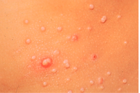
Molluscum contagiosum usually occurs in children 0-14 years of age; however, Dr Friedlander reminds us that it can be seen in adults and transmitted sexually. The highest incidence occurs in children one to four years of age. There are definite associations that we need to keep in mind, i.e, swimming and eczema both appear to be associated with either a higher risk of getting the disease or a more prolonged course.

Unfortunately, there is a subset of patients may have a more prolonged or severe course.
Screen Shot 2015-01-02 at 8.29.59 AM

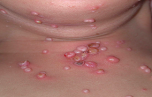
Families often want to know exactly how long it has been incubating and how long it will be there. Incubation is normally two to eight weeks. Molluscum infections can persist for years in some unlikely children, with an average duration of eight months. Generally, molluscum is asymptomatic; however, patients may be bothered with pruritus, erythema, bacterial superinfections, inflammation and pain.

What are the troublesome cutaneous findings associated with MC?

Atopy is prevalent in 44 percent of kids who present with MC (AD, asthma or allergies). What might be troublesome for you is the child who comes in with molluscum dermatitis, as this can occur in up to 39 percent of patients. The biggest problem is the patient with inflamed lesions as everyone thinks the child is infected and you need to take action. In one study, this occurred in 22 percent of patients. Another significant associated rash that we have seen is Gianotti-Crosti Syndrome-like reactions where you will see lichenoid papules around the elbows, knees, and buttocks. In children, if you note Gianotti-Crosti, type eruptions, think associated viral infections. Though we have traditionally been trained to think of EBV or hepatitis, Dr Friedlander suggests that you look to see if they have molluscum because many of them will.

What is BOTE?

BOTE is the “Beginning Of The End”, i.e., a predictor of resolution of the molluscum infection. Remember that the inflammatory phase of molluscum infection includes MC dermatitis, extreme induration and erythema, fluctuance, purulent exudate, “pseudo abcesses” and “ pseudo furuncles.” The onset of this type of inflammation to disease resolution is thought to be from three weeks to five months. (Butala N, et al. Pediatric. 2013;131:e1650.)

Why is it important to recognize “BOTE”?

The recognition of BOTE helps to avoid unnecessary cultures, antibiotics, admissions and the development of antibiotic resistance. Findings that should raise concern are a cellulitis-like appearance and lymphangitic streaking. If the child looks sick or is running fevers, you need to take action; however, if a child looks perfectly well, has multiple spots that have been red and inflamed looking for a while, and there’s no lymphangitic streaking, then you should keep BOTE in mind.

Treatment

What do the scientists tell us? Why is it so hard to clear? MC expresses proteins that circumvent immune responses (FLIPs) that inhibit b-interferon activation. What is Dr Friedlander’s hands down favorite therapy for molluscum? Cantheridin 0.7% in collodion applied with a Q-tip, washed off in four-to six hours. Some families are very nervous with this, so you can do test sites. Be sure to warn the family about the occurrence of blisters. What are the No-Nos? Catherone Plus for molluscum-NO. Do not do anything other than topical retinoids to the face , and intertriginous areas can develop severe blistering eroded reactions.

Cantharidin is an excellent treatment for molluscum but usually needs to be purchased from Canada or a compounded pharmacy. It can be ordered online through ABC pharmacy.com (Canthardin 7.5ml ~ $100.00) For those who have a willing hospital pharmacy, crystals can be ordered from Gallipot. The Cantharidin crystals themselves cost approximately $192.00/gm; when compounded it comes to $45.00/bottle.

Why don’t we let patients take Cantharidin home?

Topical application of Cantharidin for an extended time, extended areas or under occlusion and oral ingestion may cause serious side effects such as lymphangitis, TSS, and fatal poisoning. Beware of applying Cantharidin to intertriginous sites.

Another Option—The George Martin Method

  1. Obtain monochloracetic acid
  2. Fill small clean biopsy container half-way with crystals
  3. Add water half-way (saturated solution)
  4. Apply a very small amount with a toothpick
  5. Neutralize in 60 seconds with a wet paper towel

Patients can expect crusting and blistering in about two weeks.

Clinical Pearl—Remember the BOTE sign—avoid needless interventions in your molluscum patients

What do you need to think about when you see a purpuric rash?

You need to consider infection, i.e., bacterial, or viral. Also think about decreased production of marrow cells due to drug reaction, leukemia, or myeloproliferative disorders. Is there an increased consumption of marrow cells due to drug, ITP, hemolytic disorders or parvovirus? Let all of these possibilities run through your mind.

If you see lesions on the foot and hands, you may consider “hand, foot, and mouth disease” and Coxsackie. Usually Coxsackie virus is caused by A16. The disease usually occurs in spring-autumn in temperate climates and affects kids less than one to five years old. Most infections are asymptomatic. Patients may present with ulcerative stomatitis, lesions on the palms and soles and sometimes on the knees, elbows, buttocks, and genitals.

Coxsackie A6

Coxsackie 6 was first noted in 2011 by the CDC. How does it differ from regular Coxsackie? There is a wider age range with this disease. There may be vesiculobullous lesions present that are giant and a large area of involvement. There may be a rash that looks severe, but children tend not to look as sick. The problem with this disease is that if you were to try to culture it, it does not grow well in cultures. PCR however can nail it if it is available. You have to have clinical suspicion. What may help you nail the diagnosis if you don’t have PCR? Look at the mother’s nails.  And if a child comes in with nail shedding, think Coxsackie. Dr Friedlander’s practice saw several cases of onychomadesis in the office; when the moms were asked they mentioned that the children were sick about a month prior. Your first presentation of Coxsackie in the office may be a child whose nails are shedding or a mother’s.

What do we do with this child? We look at him/her, we look at the overall health of the child and we make sure that it isn’t something serious by clinical evaluation. When in doubt, evaluate for other more serious possibilities. With Coxsackie disease, we can reassure.

Look What the Wind Blew In…

A patient presents with a prolonged fever and a polymorphous eruption that looks a bit like erythema multiforme, a little bit like morbilliform, even measles-like. You need to think about bacterial and viral infections and you need to think about drug. You look at his conjunctivae and you notice limbal sparing, an area of white that is different from what you would get with adenovirus or Steven’s Johnson syndrome. When you undress the child, you notice a very distinctive perineal desquamating rash. This is Kawasaki’s Disease. You need to recognize this because while you may not see this often, when you do, you can save a life.

Kawasaki Disease (KD)

KD is a small- and medium-size artery vasculitis. There is now evidence that the incidence of KD may be linked to the velocity and direction of wind currents.

The typical age at presentation is six months to five years of age. The highest incidence of the disease is in Asian countries, and in the United States the incidence of KD is highest among the Asian population. If a child has KD, his/her siblings are six-to 30 times more at risk than the normal population and if a person had KD as a child, his/her child as a two time greater risk of the disease. The recurrence rate for the disease is two to four percent.

KD characteristics include a fever for more than five days as well as a rash that is variable but does not blister , and conjunctivitis (non exudative, non-limbal). KD affects the lips and tongue and there may be cervical lymphadenopathy as well as erythema/edema of the palms and soles as well as desquamation.

It is imperative that you intervene quickly with KD as this can decrease coronary risk by at least a factor of five.

Summary

With regards to pediatric infectious diseases, it is imperative that you know when to reassure, when to prevent unnecessary interventions and know when to act quickly!

Infectious Disease Update: Year in Review

Andrew Blauvelt, MD

Dr Blauvelt presented an update on important information and data on infectious diseases that were published in some of the top tier medical journals in 2012 and their clinical implications for the practicing dermatologist.

 Clinical Pearls
  • Look for Staph. Aureus infection in and around implantable electronic devices-these were associated with hematomas and wound healing; recommended treatment was complete removal of the device and antibiotic therapy
  • Be aware of possible bacterial contamination in antiseptic products in the office; of note, antiseptic products are NOT required to be manufactured under sterile conditions (currently under review by the FDA)
  • Erythema migrans in persons with a history of Lyme Disease most likely represents reinfection and not recurrent disease
  • Tattoos may be infected with mycobacteria contaminating the tattoo ink. Tattoo ink does not have to be manufactured in sterile conditions. M. Chelonae was found recently in a batch of tattoo ink. Diagnose, treat, and report accordingly
  • Travel history is important for a sick patient with cutaneous abscesses.  Meloidiosis, a disease caused by environmental (in soil) gram-negative bacillus Burkholderia pseudomallei, presents with multiple cutaneous abscesses and pneumonia. It has a 40% mortality rate.
What about viruses?
  • Adults born from 1945-1965 should receive one time testing for the hepatitis C virus without prior assessment of HCV risk (recommendation from the CDC)
  • All persons identified with HCV infection should receive brief alcohol screening/intervention as indicated followed by an appropriate referral to a specialist
  • Clinical data and ex vivo experiments that show pre-exposure prophylaxis with anti-HIV drugs can block subsequent infection; however, controversy exists over ethics, practicality, and cost
  • Genital herpes simplex virus 2 (HSV-2) enhances sexual transmission of HIV
  • Antiviral medication for the treatment of HSV-2 using short courses of high-dose acyclovir and valacyclovir (1 gram TID) may decrease outbreaks, but does not prevent  subclinical HSV-2 genital shedding of the virus.  The therapy does decrease shedding by about 50% but the rate of breakthrough shedding was 16-20 episodes/year. Novel therapies need to be developed to block shedding
More Clinical Pearls
  • Live vaccines may be safer in the setting of biologic therapy than previously believed.  In a study of MCR patients receiving biologics, 633 patients were inadvertently vaccinated with the herpes zoster vaccine. None developed varicella or zoster.
  • In addition to previously identified in EVER1 and EVER2 genes, epidemodysplasia verruciformia may be caused by a genetic defect in RHOH which leads to T cell defects
  • Injectable corticosteroid products may be contaminated during the compounding or manufacturing process
  • Natural disasters can lead to unusual medical conditions.  Mucormycosis, caused by infection with ubiquitous molds in soil and decaying wood, resulted in 13 cases of mucormycosis resulting in 5 deaths in storm affected areas. The median number of wounds through which the mold penetrated was 5.
  • Bilateral leg lymphedema, which can lead to elephantiasis verrucosa nostra, is associated with obesity (and not filariasis in non-endemic areas).  The average BME was 51.4.
  • Topical 0.5% Ivermectin lotion is a promising new therapy for head lice. In two large studies, a single application of 0.5% ivermectin applied to dry hair, left for 10 minutes, and then washed off resulted in nearly 75% of patients being louse free at day 15.  Nit combing in not necessary because ivermectin if both pediculocidal and ovacidal.
Aditya Gupta, MD, PhD

Dr Gupta, one of the leading authorities in the world on fungal infections, provided the audience with an update onychomycosis and how dermatologists can improve upon the management of this condition.  Dermatologists should be aware of the prevalence of onychomycosis and this may help practitioners understand why patients may fail certain therapies. There are various therapies available for onychomycosis including oral therapy, topical therapy, and devices.

Oral Therapy

Terbinafine is probably the gold standard for the management of onychomycosis. Dr Gupta and his colleagues studied various terbinafine dose regimens and found that 2 pulses 250mg/day 4 weeks on/4 wks off/4 wks on appeared to have the best outcomes; however, there was not a significant difference between continuous and pulsed regimens.  It is important that dermatologists understand the existing data on the various antifungal medications currently available in order to apply this information into clinical practice.

Topical Therapies

The nail plate is the greatest barrier when treating fungal infections. Currently, ciclopirox is the only topical therapy approved in the United States. However, there are emerging therapies currently under investigation.

Device Therapy

There are lots of questions about laser therapy. Podiatrists seem to use laser therapy quite often; yet, dermatologists continue to raise questions about their effects and how they actually work. Laser therapy poses the same challenges as those of topical therapies. To date, there is only one published trial (n=13) and the study demonstrates an efficacy rate of 51%. More studies are needed to determine if laser treatment is effective.

In conclusion, dermatologists need to recognize the importance of the choice of agent for onychomycosis and identify other important management strategies that may help to improve the outcomes of their patients.