Cutaneous Oncology: Clinical Pearls

George Martin, MD & Ted Rosen, MD

Why field therapy?

Data from Europe using a variety of techniques (including full face cross-polarized light examination, fluorescence photography, high definition optical coherence topography and reflectance confocal microscopy) clearly validate the concept of a “field” of abnormality in close spatial proximity to visible AK lesions.

Facial AKs:

Although we did not mention this in our talk, for those of you who use 5-FU, please remember that the Phase III data on 0.5% 5-FU demonstrated that 1 week of daily use of 0.5% 5-FU cleared nearly 75% of individual AKs. Try: 0.5% 5-FU QD x 1 week, wait 1 month, then follow with 2 -3 weeks QD application to “clean up” remaining AKs. This regiment has gained widespread acceptance by patients and physicians as a more tolerable field therapy. 5% 5-FU BID is equivalent to 0.5% 5-FU and can be used interchangeably.

Ingenol mebutate 0.015% applied nightly x 3 has been a remarkably effective therapy with great patient compliance. However, it’s FDA approval was for limited areas (25 cm2). As a full-face therapy, it appears very effective; however, controlled studies on “full face” clearance/efficacy is pending. Patients need to be counseled that they will experience a moderate to severe “sunburn-like” effect beginning 4 hours after application. This is likely due to its MOA, which includes a direct cytotoxic effect. Analgesia is generally required. Clinically, we have observed while treating full-face, ingenol mebutate is selective for AKs with mild/moderate erythema between AK lesions.

New data, both short term (11 weeks) and long term (one year), support the benefit of using ingenol mebutate in combination with standard liquid nitrogen cryotherapy. Cryotherapy is performed first, and then ingenol mebutate used per approved protocol three weeks late. At one year, the combination therapy has a higher complete clearance rate than cryotherapy followed by vehicle control. Off label, ingenol mebutate appears promising for the management of actinic cheilitis in the three day, FDA-approved regimen. More studies are needed to validate this idea.

If you perform PDT in your practice, and use 1 -> 3-hour ALA incubation periods, recent Phase II studies from DUSA show that 1, 2 or 3 hour incubation periods are roughly equally efficacious (35% – 50% clearance) but 2 treatments 8 weeks apart are required for most patients to achieve >70% individual lesion clearance. To maximize the efficacy of a 1-hour incubation, consider pre-treating with 5-FU for 1 week to the face or 10 days to the scalp then perform a 1 hr. ALA incubation. This combination will eliminate the need for a 2nd PDT. For those patients with refractory facial AKs, consider pretreating for 7 days with 3.75% imiquimod followed by ALA PDT (1-3 hour incubation). Excellent long-term results (18 months) have been observed when destructive techniques such as PDT are combined with immune modulators.

Can ALA PDT be “painless”?? Try incubating for 15 mins with ALA and then place the patient under the blue light for 1 hour. Preliminary results (G.Martin MD) demonstrate that ALA PDT as monotherapy or in combination with 5-FU or 3.75% imiquimod, employing this technique is in fact “painless”. Individual AK clearance rates of 50% were demonstrated in a proof of concept small patient study. Large-scale studies are warranted to determine efficacy. Network meta-analysis (comparing different techniques against one-another) suggest that PDT is the optimal field therapy for AKs.

The focus of PDT has been aimed at treating AKs. Is there evidence that PDT may prevent BCCs? Data from studies on basal-cell nevus syndrome patients demonstrate long-term clearance and prevention of new BCC development compared to non-treated areas of the trunk following ALA PDT using red light.

The use of 3.75% imiquimod for diffuse facial AKs while effective, results in substantial “downtime” of nearly 6 weeks. Consider 3.75% imiquimod QD x 7 days, 2 weeks rest, followed by once weekly applications. There will be some initial unsightliness during the 1 week of continuous use. Individual AKs are seen to clear with continuous application with minimal irritation. Chronic immune stimulation (>1 year) appears to be helpful in limiting AK recurrences and may prove over time to inhibit the development of invasive SCC. Large-scale studies are warranted.

New Drugs and New Concepts: Clinical Pearls

Neal Bhatia, MD & Ted Rosen, MD

  1. New drug for papulo-pustular rosacea: QD 1% ivermectin cream (Soolantra®, 30g tube)
  • Better than placebo and superior to metronidazole 0.75% BID
  • “Clear” or “Almost clear” by IGA : 38-40%
  1. New HPV vaccine (Gardasil 9®)
  • Contains VLP to prior quadrivalent vaccine (HPV 6,11,16,18)
  • PLUS: contains VLP to immunize against HPV 31,33,45,52,58
  • Now 97% protective against genital SCCA due to 90% etiologic HPV
  • Also recommended for MSM, where ~75% protective against anal SCCA
  1. Miltefosine (Impavido®) for leishmaniasis
  • Both old world and new world organisms (more evidence for new world)
  • 100-150mg daily (higher dose if over 45kg weight)
  • AEs: anorexia, nausea, vomiting, diarrhea, H/A, mild ↑ LFTs, mild ↑Cr, and mild thrombocytopenia; but is Pregnancy category X (contraindicated): Do not take if pregnant, use adequate contraception during Rx and for five months after therapy has been discontinued
  1. Biologics are coming for atopic dermatitis
  • Duplilumab (anti IL4, IL13)
  • Lebrikizimab (anti IL13)
  • Mepolizumab (anti IL5)
  • Various anti IL31 monoclonal antibodies
  1. Watch for Vitamin D deficiency in various derm diseases: psoriasis, SLE, hidradenitis, alopecia areata; Not known if repletion of Vitamin D will be therapeutic
  2. Tofacitinib, a JAK3 janus kinase inhibitor, currently approved for RA, may be effective in psoriasis and psoriatic arthritis. A case report appeared showing benefit in alopecia totalis!
  3. Adalimumab appears beneficial for hidradenitis in a Phase III RCT.
    • Dosage 40mg weekly
    • Still not a miracle drug
  4. Tonsillectomy improved refractory psoriasis!
  5. Red henna tattoo does NOT affect pulse oximetry
  6. RUSHING to put on condoms leads to errors and failures, including increased STDs

Malpractice and Informed Consent

Mathew M. Avram, MD, JD

One of the keys to a happy patient is to set realistic expectations and to obtain an appropriate informed consent. Dr Avram, a world-renowned cosmetic surgeon and former practicing attorney, provides us with some important basic information and key concepts that can help us navigate some potentially confusing legal issues.

There are four required elements to a malpractice suit:

  • Breach
  • Of a duty
  • That causes
  • Harm

Duty

The standard is that of a “reasonable duty.” This is the quality of care at a level consonant with medical knowledge and judgment that a physician is reasonably expected to posses. When that standard is litigated, there are a couple of different ways that this can be done, e.g., expert testimony, medical literature, standard practice, and clinical guidelines. When using expert witness qualifications, remember that the expert must have sufficient training to assess the physician’s performance. Keep in mind; however, that this is ultimately at the trial judge’s discretion and there is no requirement for board certification.

The plaintiff’s expert testimony must establish a standard of care and must be able to establish that the physician failed to meet that standard to a “reasonable degree of medical and scientific certainty.”

Negligence can also be established through:

  • Cross examination of defendant’s expert witness
  • FDA/PDR warnings
  • Learned treatise
  • Defendant admission of negligence—“Sorry, I hurt you. Don’t worry, I have insurance.”

Defenses to a Malpractice Suit

Respectable Minority Exception

This is where there are two (or more) schools of thought as to a procedure and a physician can pursue one of many treatment options that a “considerate number of reputable and respected physicians” pursue. (E.g. cryotherapy versus topicals for AKs)

Contributory Fault

This is where a patient makes mistakes or certain lifestyle choices. Damages to the plaintiff are mitigated by failure to exercise ordinary care, i.e., drinking alcohol while taking terbinafine.

Statute of Limitations

These are usually two to three years after injury or malpractice. The key is the timing, i.e., the timing from when the defendant breaches his/her duty or when the plaintiff suffers injury. What can make this complicated is when the plaintiff becomes aware or reasonably should be aware of the injury. (e.g., delayed hypopigmentation six months after CO2 resurfacing or granuloma years after a permanent filler treatment) Depending on the procedure and its consequences, the statute of limitations can be extended.

Doctrine of Informed Consent

Remember that your informed consents are NOT going to protect you against a malpractice case and vice versa. These are two distinct legal theories by which you can be sued.

All patients have the right to an informed consent prior to any treatment.

Failure to obtain an informed consent constitutes a battery and the physician is liable for civil damages. Consent can be written or oral.

As a physician, you need to examine the probability of risk and the severity of the side effect. Thus even the small chance of blindness with soft tissue filler injections needs to be included in your consent forms. You also need to examine particular susceptibilities of the patient, i.e., hyperpigmentation in a patient with a darker skin type undergoing laser procedure. Your duty becomes expanded as the patient asks more questions.

In terms of your consents, keep it simple. A written consent is ineffective if the patient doesn’t understand material information about the procedure. Avoid technical medical terms and use lay person terms.

Purpura–Bruising

Erythema–Redness

Standards of Disclosure-The patient must have the capacity to make a medical decision. Remember that minors lack this capacity and parents must consent to all medical or cosmetic procedures.

Blanket Authorizations-Overbroad written consents are viewed by disfavor by courts, they like more specificity to show an informed consent.

If a patient misleads a physician, this can be defense to an informed consent action. The patient has a duty to disclose accurate information. Regarding patient decision making limitations, absent of showing incompetence, the courts presume patient comprehension.

Tips to Avoid a Law Suit

Patient selection is a key decision. Assess the patient the patient at consultation, particularly their expectations. Trust your own intuition about a patient as well as your staff’s intuition. For elective, cosmetic treatments, don’t be afraid to say NO.

Studies have shown that your relationship with the patient may be the single most important factor in avoiding a lawsuit. Patients with a good relationship with their physician are far less likely sue.

Avoiding Complications

Know your limits—even in skilled hands, if you treat a sufficient number of patients, you will encounter challenging side effects. Do not perform a procedure that might produce a single side effect that you cannot recognize and treat. Remember the complications are inevitable; however, good practice and common sense can minimize complications significantly. Do not abandon or avoid a patient with a poor outcome or side effect. This is bad medicine and rightfully angers your patients increasing your risk of a lawsuit.

If you are uncertain as to what is happening, you should consult with a colleague. Temporary and expected side effects are common. These include erythema, edema, and purpura. Legal consequences are less likely with adequate patient handholding.

Regarding botulinum toxins, litigation for cosmetic treatments is rare and less common than for lasers. With lasers we may see temporary pigmentation, but time is on your side and the discoloration will most likely resolve before any litigation proceeds.

More permanent side effects are more likely to produce liability (e.g. scars, blindness). It is important to distinguish how long-term these side effects actually are.

Documentation

Documentation is crucial. Document physical findings on the exam, even the ones that aren’t necessarily related to what’s happening. Good photography is important because poor photography will work against you. Also ensure that your consent forms are properly filled out.

Lasers and Lawsuits

A study by Dr Avram and his colleagues published in JAMA dermatology looked to identify common errors/risk factors for litigation in laser surgery through a search of an online legal research database. They reviewed over 1600 documents that were retrieved by their search criteria and found 182 unique legal claims involving injury from a cutaneous laser treatment. Not surprisingly, New York, California and Texas were the most common areas, given their populations. The most common litigated procedures were hair removal, rejuvenation (mostly IPL), and vascular treatments. The most common injuries sustained were burn, scar and pigmentation. The most common causes of action were standard of care, informed consent, and fraud (exaggeration of benefits).

Non-Physician Litigation Data

From 2008 to 2011, there has been a dramatic increase in litigation against non-physician operators. In 2008, non-physician laser treatments and litigation represented 36.3 percent of all laser-related cases whereas in 2011 it represented 77.8 percent of cases. Only 23.4 percent of non-physician operation litigation cases arose in medical office settings. 76.6 percent of the cases were performed outside a traditional medical setting such as salons and spas (mainly laser hair removal). The majority of these cases were performed without any direct physician supervision.

Physician Extenders

The physician is held liable for a physician extender’s negligence provided that he/she is an employee receiving a salary, benefits and performing within his/her scope of duty. This is regardless of whether or not the physician saw the patient at the time of the visit. Supervision of non-physician laser procedures varies from state to state. Some jurisdictions require on-site supervision by physicians, while others do not.


The Future of Laser Dermatology

Rox Anderson, MD

Dr Anderson, a professor at Harvard Medical School in Boston, provides us with his views on the future of lasers in dermatology. A lot what the future holds for us is going to be driven by different motivators. Economic change, both globally and nationally, is one of these factors. Dr Anderson feels that we have an obligation, not only for society and our patients, but also to ourselves. Similar care at a higher cost is no longer going to be tolerated. The current generation of dermatologists must embrace and take control of who “extends” them, and how. We have to focus on simplicity, access, efficiency, the idea of being consumer-friendly, and new technology such as apps and social media/crowd-sourcing. We need to be able to provide more for our patients, but we also need to be better and cheaper. What are our “sacred cows?” Who will pay? Who will take the hit? We have to look at what we’re doing and stand back to see how we can be doing it differently.

Dr Anderson predicts a dozen big changes in the field by 2025:

  • Smarter lasers that can help reduce risk and monitor skin pigmentation
  • Fully software-programmable laser targeting
  • A cadre of prescription home-use smart laser, IPL, US devices—all we have right now is basically the hair removal devices
  • Melasma will be figured out à specific, effective topical therapy
  • Same-day laser tattoo removal, reliably, without scarring
  • Useful live microscopy (the real question is, who will read it? what are they used for?)
  • Microbiopsies à rapid tissue diagnostics without the pathologist
  • “Mohs for Schmohs” –if you can accept the idea that you can have microscopically-guided laser surgery, this will be a video game that the millennials will love playing
  • Smart-phone dermatology apps: from ~2000 now to ~200,000
  • Excellent, automated melanoma detectors where they belong
  • Acne will, in essence, be cured
    • Devices that “target” sebaceous glands, ala hair removal
    • Sebum inhibitors other than retinoids: PDT, new drugs
  • Sunscreens that never wash off

Acne

The one thing that oral retinoids have going for them is that they are the only medicine we have that have a potent suppression of sebum, i.e., sebaceous gland function. What about other ways to do that? If you look at the microanatomy of skin, it is just another part of the hair follicle and Dr Anderson believes that there are many other strategies that can be used to do selective injury to those glands.

Screen Shot 2015-01-12 at 7.47.35 AM

If you look at the absorption spectrum of sebum, at 1726 nm the sebum has a little more absorption than water. This has to do with its lipid content. Dr Anderson and his colleagues used a giant, free electron laser to conduct an experiment at that wavelength testing this hypothesis—whether sebaceous glands can be selectively injured with photothermolysis. The answer is yes. (Sakamoto FH, et al. Selective photothermolysis to target sebaceous glands: theoretical estimation of parameters and preliminary results using a free electron laser. Lasers Surg Med. 2012; 44(2):175-83,) Dr Anderson has a high-powered benchtop prototype 1726 nm laser in the lab specifically designed to go after sebaceous glands…stay tuned.

Sebaceous cryolysis—cold-induced selective injury, could this apply to sebaceous glands in the same way it has been used for fat removal? We know that the sebaceous glands are loaded with lipids, just like fat. At the Wellman Center for Photomedicine (an MGH research center directed by Dr. Anderson) Drs. H.Ray Jalian and Joshua Tam and Conor Evans have been studying the biologic consequences to the sebaceous gland of controlled cold cycles. We see the same things that go on in adipocytes, and actually it’s much easier to trigger because the glands are more superficial.

Dr Anderson feels that we will end up with a cold-induced treatment for acne.

Another strategy for acne would be to put something in the gland that absorbs light. One of the issues with acne drugs is that they are tough to get into the gland. The idea here is to apply something topically, force it into the sebaceous gland, and “fry” it with a pulse of light. In one study, core-shell microparticles worked the best. If you take a nanoparticle and make it out of either silver of gold, plasmon resonance occurs. Silver and gold are both noble metals that have very high conduction so the electrons in them are very free to move. We have an incoming optical field and at the right frequency it is in resonance with the conduction band electrons providing a tremendous amount of energy. You can also design the particles to have their own absorption.

Acne Treatment With Gold-coated Particles

  • Clean skin
  • Apply particles topically
  • Massage to deliver them into follicle
  • Remove residual from skin surface
  • Treat with laser at the absorption peak wavelength, combined with skin surface cooling

Does this work? Yes, it does.

Using two-photon microscopy in a collaboration with James Tunnell (UT Austin), it was shown that gold particles can be forced into sebaceous glands. After exposure to an 800 nm diode laser, one can see local destruction of sebaceous glands.

Screen Shot 2015-01-12 at 7.47.50 AM

Microscopy and Smart Lasers

Optical coherence tomography is a means of microscopic imaging. It is fully deployed in ophthalmology and has been for over a decade. In dermatology, it’s used mostly as a research tool.

Screen Shot 2015-01-12 at 7.48.01 AM

You can see beautiful structure—epidermis, sweat ducts, vasculature, etc. but this hasn’t made its way into clinical practice. If you look further into advanced laser microscopy, we can do some very interesting things. Coherent anti-Stokes Raman scattering (CARS) microscopy requires no stains. It’s a way of setting up a laser microscope that excites the vibrational modes of bonds in various molecules. The bonds are quite specific to the molecule, so you can look at molecular species in the skin. You can tune this process to look at DNA, protein, and lipids. Of great interest, if you replace hydrogen with deuterium, the carbon-deuterium bond is in a very quiet region of the spectrum. You can label drugs with deuterium quite easily and it’s non-toxic. The picture on the bottom right is the first picture that Dr Anderson has ever seen of directly imaging a drug uptake into human skin in a living state. This was performed by tracking deuterium as a label.

Screen Shot 2015-01-12 at 7.48.10 AM

The lipid imaging by CARS also gives us beautiful pictures of sebaceous glands. The image above shows sebaceous glands in living mouse skin.

Another major advance, would be to have in vivo microscopes that yield images similar to the familiar H&E staining of conventional histopathology. Dr Daniel Gareau at the Rockefeller Institute has been doing work with digitally stained confocal microscopy. (D.S. Gareau, J. Biomed. Opt. 2009.14(3).)

Screen Shot 2015-01-12 at 7.48.22 AM


Screen Shot 2015-01-12 at 7.48.33 AM


If you look at the reflectance, it shows you where the fibers of collagen are. By staining the fluorescence and the reflectance different colors and putting them together, it gives us something that we, as dermatologists, have all been trained to look at. We are going to see his technique, along with other ones, being melded into a user-friendly version. This entire process takes three minutes and doesn’t require any technicians.

Screen Shot 2015-01-12 at 7.48.42 AM

Eulerian Imaging

Remember that except at absolute zero, everything moves. The minor, small motions are not visible; therefore, we think things are stationary. Professor Bill Freeman (MIT) has recently developed Eulerian imaging. It detects small changes in position or color, amplifies them then adds them back onto the original video image. Skin hemodynamics can be revealed in detail, using nothing more than your smart phone. Dr Anderson believes that we may see apps in the future for dynamic oxygen saturation, apps for motile cells, and apps where we can measure the rate of cancer growth. To view Eulerien imaging of the skin, visit: http://www.youtube.com/watch?feature=player_embedded&v=e9ASH8IBJ2U

A New Sunscreen that even Dr Anderson will Use!

There is a new sunscreen under development in collaboration with MIT, Harvard/MGH, and Living Proof. This sunscreen is wearable, breathable, invisible, elastic, and lasts all day. This was first used cosmetically, but it turns out that this film has other properties. What’s interesting is that this film has an SPF of 15 and can be worn for days.

Things That Sound Impossible Now, by 2050:

  • Real rejuvenation
  • Skin as a user interface
    • Synthesis &/or release of drugs or proteins on demand
    • Activable implants: particles, designer cells, machines
    • Biosensing tattoos and other monitors
    • Light-emitting skin
  • Hybrid skin: mixing it up with other organs
  • Magic Wands

The Magic Wand Project

Every year, Dr Anderson makes just one New Year’s resolution. His 2013 resolution was to somehow empower the young clinicians to actually lead research. Using a 3-D printer, Dr Anderson made some magic wands and gave one to each of the young dermatologists in his department. His instructions were that anytime he/she faced a problem in clinical practice that couldn’t be solved, point the wand at the problem and write it down. Most clinicians do not reflect upon the unique potential that they have, to motivate biomedical research. No one else understands the problems the way that clinicians do. You can’t really define the problem unless you really understand it. Among the clinicians at Mass General, they used the magic wand for one month in practice and generated a list of about thirty practical problems in dermatology that are worth solving. Out of the thirty problems, eight active research projects are underway; each led by a clinical dermatologist.

  1. Reliable diagnosis of cellulitis
  2. The world’s best ever sunscreen
  3. A much better treatment for warts
  4. Long-lasting blockade of itch and pain
  5. A great treatment for onychomycosis
  6. Correction of field cancerization
  7. Medical grade, personalized cosmetics
  8. KS / HHV-8 diagnosis in Africa

One of the challenges that we face is to bridge the gap between clinical practice and scientific research.  Dr. Anderson enjoys trying to do that.


MauiDerm News Editor-Judy Seraphine


Systemic Agents

John Zone, MD

Dr Zone, an expert in systemic disease and Co-Director of the Immunopathology Lab at the University of Utah, discusses the medications that we use to treat these various conditions. Most of the therapies discussed in this presentation are off-label unless otherwise noted.

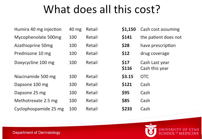
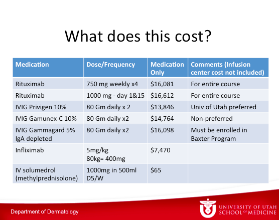
What does all of this cost? The table below depicts the numbers from the University of Utah pharmacy.

Screen Shot 2015-01-06 at 7.05.21 AM

Dr Zone has said this before, if he gets pemphigus, he wants to take rituximab. He is convinced that rituximab is not only the best treatment, but it is the cheapest in the long run.

Screen Shot 2015-01-06 at 7.05.44 AM

IgE

IgE is more under the purview of the Allergist, so it isn’t discussed much in dermatology. It is a monomer and is present in very low concentrations in the body. Its synthesis is 25 to 2000-fold less than other immunoglobulins making it difficult for us to measure. It lasts in the blood for 48 to 72 hours so it has a very short half-life. IgE doesn’t activate C’ and instead binds on mast cells for weeks to months. If you develop a sensitivity, once the IgE molecules bind to mast cells, they are going to be there for awhile no matter what you do.

The approach that we take to treat conditions like urticaria always astounds Dr Zone. We know that IgE binds to mast cells in our skin. When the mast cell becomes degranulated, it releases histamine, TNF, proteases, and heparin—the things that cause people to get symptoms of itching and swelling manifested as urticaria. We treat people with antihistamines– we give people something to block the effect of the histamine after it’s released. There are a number of papers studying the positive effects of TNF inhibitors in urticaria. At Dr Zone’s institution, they have treated about thirty people with fairly good results. If you have intractable urticaria, you are open to a lot of things. Over a longer period of time, there is release of prostaglandins and leukotrienes. There are prostaglandin inhibitors and montelukast that can be used as well for urticaria. Over the long run, we then see eosinophil recruitment and release of other cytokines.

Given this information on pathogenesis of urticaria, it is clearly better to block the effect of IgE and prevent the end results?

Screen Shot 2015-01-06 at 7.06.05 AM

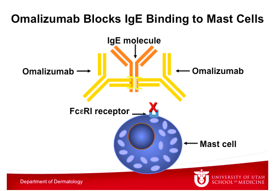
This graphic illustrates Omalizumab binding to an IgE molecule, which also takes place at the constant region of the IgE molecule. Note that IgE binds either to the FceRI receptor on the mast cell or to Omalizumab, but it cannot bind to both at the same time. Omalizumab inhibits the binding of IgE to the high-affinity IgE receptor FceRI on the surface of mast cells and basophils. Reduction in surface-bound IgE on FceRI-bearing cells limits the degree of release of mediators of the allergic response. Treatment with omalizumab also reduces the number of FceRI receptors on basophils in atopic patients.

Omalizumab is approved for asthma at this time and has recently been approved for refractory urticarial. To read more about omalizumab, the following references may be helpful:

  • Pemphigoid
    • G Ital Dermatol Venereol. 2012 Jun;147(3):251-7.
  • Urticaria
    • Curr Opin Allergy Clin Immunol. 2012 Aug;12(4):406-11
  • Atopic dermatitis
    • Clin Exp Dermatol. 2012 Oct 22

There are going to be a lot more studies on urticaria and atopic dermatitis with omalizumab. Dr Zone uses Omalizumab 150-375mg subcutaneously every two to four weeks. It costs anywhere from $500 to $2000 per month and he has been able to get insurance companies to cover it. Regarding side effects, anaphylaxis has been reported; however, omalizumab is rather well tolerated.

Intraveneous Immune Globulin in Autoimmune Diseases

IVIG is currently used for the following dermatologic disorders:

  • Pemphigus vulgaris
  • Pemphigus foliaceus
  • Bullous pemphigoid
  • Mucous membrane pemphigoid
  • Epidermolysis bullosa acquisita
  • Toxic epidermal necrolysis
  • Necrotizing fasciitis

There is mixed information suggesting that IVIG works; but because these diseases are very severe, there is likely to be approval.

IVIG comes from thousands of donors and there is no data to show that one brand is better than another. Somewhere around one percent of the population are IgA deficient. If you are IgA deficient, you can have an anaphylactic reaction when IgA-containing immunoglobin is administered. Dr Zone believes that everyone should have a total serum IgA done before receiving IVIG. If you cannot do this, then the patient should be given the IgA depleted therapy. IVIG is used in smaller doses for immunodeficiency.

The therapeutic effects of IVIG most likely reflect the function of natural antibodies in maintaining immune homeostasis in healthy people. We think that antibodies are just for killing bacteria and viruses, but it’s not just that. Antibodies regulate our immune system. When you get someone else’s IVIG, you have a great chance of changing the regulation of your immune system.

The debate on toxic epidermal necrolysis (TEN) and IVIG continues. Ten or so years ago, Dr Zone was convinced that IVIG was the best treatment for TEN; but now he’s not totally sure. Huang and colleagues conducted a systematic review and meta-analysis looking at the efficacy of IVIG on the treatment of TEN. Seventeen studies were included. (Huang, et al. BJD 2012:167:424-432) The studies reported that pediatric patients had lower mortality than adults and adults with high dose IVIG had lower mortality compared to low dose IVIG (18.9% vs 50%). However, logistic regression failed to show a significant correlation with mortality.

What would Dr Zone do if he had TEN? After studying the papers, he would take IVIG.

IVIG for SJS/TEN Overlap and TEN:

  • Start IVIG 1 gram per kg for 3 days or 0.75 gram per kg for 4 days
  • If there is any concern for IgA deficiency, start IgA depleted IVIG until IgA levels are confirmed
  • In cases of renal failure, obtain renal consult and consider 0.5 gram per kg for 4 days vs. short course, high dose steroids

Dr Zone believes that the survival is better if it is done carefully. He would also most likely take IVIG with progressive Stevens Johnson’s Syndrome. IVIG has been used for over thirty years for patients with primary or secondary antibody deficiency and has not been found to have long-term side effects. IVIG has been tested by PCR for hepatitis B and C and HIV. There have been no cases of HIV transmission and no recent cases of hepatitis transmission. Side effects of IVIG include:

  • Headache, migraine
  • Fatigue
  • Anaphylaxis
  • Aseptic meningitis
  • Congestive heart failure
  • Pulmonary edema
  • Acute renal failure
  • Leukopenia
  • Hemolytic anemia
  • Hepatitis
  • Venous thrombosis

There are a group of people who believe that cyclosporine works for SJS/TEN. A study of 29 patients was published in the British Journal of Dermatology suggesting a possible usefulness of cyclosporine in SJS and TEN that needs to be confirmed. (Valeyrie-Allanore L, et al. Br J Dermatol. 2010;163(4):847-853.)

Takeaway point—If Dr Zone had SJS/TEN, he would take IVIG. If you didn’t want to give IVIG, cyclosporine is an option. Otherwise, you are in for a disease with a thirty percent mortality rate.

Scleromyxedma

This is a very rare disease and as dermatologists, we will likely see one case in our lifetime. A case series from Johns Hopkins reported eight of out ten patients with either 100 percent complete or partial response rate with IVIG treatment 2G/kg/month in divided doses. In the old days, there was no good treatment for scleromyxedma. Once again, if Dr Zone had scleromyxedema, there is no doubt that he would take IVIG. You do end up taking it indefinitely, but we try to decrease the dose and frequency.

Oral Corticosteroids

Dr Zone feels that he sees a lot more side effects from oral corticosteroids than immunosuppressive therapy. Before starting a patient on oral corticosteroids, it is important that you check their blood pressure as well as glucose, electrolytes and creatinine. Dr Zone gives H2 blockers or proton pump inhibitors to most people who are on therapy for more than two or three weeks. Gastritis and upper GI bleeding is a common side effect. For more than two or three weeks, Dr Zone also uses osteoporosis prevention. It is also important to monitor side effects every two weeks.

How can we prevent osteoporosis?

  • Suggest Calcium carbonate plus D for short term (twice daily)
  • Bisphosphanates
    • Alendronate 5 mg daily or 35 mg. weekly
    • Beware of esophagitis
  • Calcitriol (Vitamin D3)
    • .5-1.0 micrograms daily
    • Beware of serum calcium
  • Estrogen or Testosterone

You can start prednisone at approximately 1mg/kg/day. The patient can take the entire dose in the morning or BID early in the day. Alternating day therapy prevents adrenal suppression, but not osteoporosis. It is extremely important that you try to control the disease then minimize the dose, not the reverse. The reason that you want to taper steroids is to avoid rebound in the short term and manage adrenal suppression in the long term. If you want to evaluate for adrenal suppression, you can do an 8am cortisol and ask the patient not to take their steroid.

Dapsone

Dr Zone uses a lot of dapsone in his practice because he treats a lot of patients with dermatitis herpetiformis (DH). Dapsone inhibits neutrophil chemotaxis and attachment. It has neither an effect on antibody or complement deposition nor an effect on the reaction to gluten in DH. Dapsone simply blocks the inflammation in the skin.

Every patient on dapsone gets hemolytic anemia. Dapsone is a sulfone, meaning it has a double-bond oxygen—an incredible oxidant stress. What does that do to red cells? Red cells older than 90 days don’t have much G-6-PD because it decreases as cells age. They don’t have the ability to withstand oxidant stress. When you give dapsone, you’re going to hemolyze cells that are over 90 days old. If the red cell lifespan is 120 days and you give dapsone, you might hemolyze one quarter of the red cell mass. What does this mean? If you’re a young person you might feel a little fatigued; however, if you’re 80 and have congestive heart failure (CHF), it might end up exacerbating the CHF. But remember that everyone is going to get hemolysis. What happens? Your bone marrow starts to produce reticulocytes so all of a sudden the mean cell volume (MCV) goes up. When you treat someone with dapsone, you will see macrocytosis (because of the big reticulocytes) and a slight fall in the hematocrit, or you may see a fairly big fall in the hematocrit. With time, most people will compensate enough so that they’ll have enough reticulocytosis that they’ll raise their hematocrits back to the normal level. They’ll still be constantly breaking apart the red cells that are more than 90 days old because they can’t withstand oxidant stress.

Before giving dapsone, you should perform a baseline CDC and chem profile as well as a G-6-PD in Asians, African Americans, or those of southern Mediterranean descent. Start dapsone at 25mg daily and increase by 25mg weekly until the symptoms are under control. A CBC should be done weekly for the first four weeks, then monthly for six months, then semiannually. A chem profile should be performed at six months and then annually.

Dr Zone has treated somewhere around 1000 people with dapsone. He’s had two people who have developed leukopenia within two weeks. You can get aplastic anemia that will occur within the first four to six weeks. This is why you want to check the blood counts frequently in the beginning.

Mycophenolate mofetil (MMF)

MMF was approved by the FDA in 1995 for the prophylaxis of acute rejection in renal transplant patients. It is an ester of mycophenolic acid (MPA) and is synthesized to increase the bioavailability of MPA. It works by inhibiting leukocyte recruitment and glycosylation of lymphocyte glycoproteins involved in endothelial and intercellular adhesion and lymphocyte trafficking.

When you think about what we do as dermatologists whether it’s eczema or severe dermatitis, we are treating lymphocytes. MMF is a fairly good to drug to use in these cases. It’s effective as a corticosteroid-sparing agent in corticosteroid responsive dermatoses. MMF takes six to eight weeks to have an effect. You should start patients at 500mg bid. The most effective dose is 1000mg bid and the maximum dose is 1500mg bid. You may want to monitor levels if you are concerned about toxicity.

Dr Zone has seen the following side effects in patients receiving more than 2 g/d:

  • GI disturbances (nausea, diarrhea, dyspepsia, abdominal pain)
  • Persistent cough
  • Reversible hematologic side effects

It is unknown as to whether or not MMF increases the risk of lymphoma and carcinogenicity. There is an increased risk of viral and bacterial infections.

Azathioprine

Dr Zone uses a lot of azathioprine and feels that it’s a great drug. Everyone should get a thiopurine methytransferase (TPMT) level before starting azathioprine as well as a CBC and comprehensive metabolic panel. Azathioprine is converted to 6 mercaptopurine within one hour. It works by inhibiting T lymphocyte function and Ig synthesis.

Dr Zone starts at 50mg/day and increases to 3mg/kg/day over one to two months. Some people get severe nausea and/or an acute febrile reaction. With severe nausea, most patients just can’t take the medication. With mild nausea, you can divvy up the dose to three times per day. It is important to monitor CBC for leukopenia and perform liver function tests for obstructive patterns and hepatocellular damage. You can taper steroids and continue the azathioprine.

Cyclosporine

Here’s the trick with this…the pharmacy may call you and ask if you want to use “modified” or “non-modified.” “Modified” cyclosporine (gengraf and neoral) has more consistent absorption than non-modified (sandimmune). They are NOT bioequivalent. It is important that you use one and stick with it. Before starting cyclosporine, you should perform a baseline CBC, metabolic profile and check magnesium.

Start cyclosporine at one to two mg/kg/day. Remember that hypertension and elevation of creatinine are limiting factors. Leukopenia may occur as well as hypomagnesemia. You should monitor CBC and metabolic profile monthly. Patients can also monitor their blood pressure at home.

Cyclophosphamide

This is used for ocular cicatricial pemphigoid and refractory bullous diseases as well as refractory vasculitis. If you haven’t used a lot of this drug, you should probably refer the patient to someone with experience. Cyclophosphamide can cause leukopenia as well as alopecia and gonadal damage. There is also a risk of leukemia and bladder cancer.


Infectious Disease Update: A Tale of Three Rashes

Sheila Fallon-Friedlander, MD

Molluscum Contagiosum (MC)

Screen Shot 2015-01-02 at 8.29.38 AM

Molluscum contagiosum usually occurs in children 0-14 years of age; however, Dr Friedlander reminds us that it can be seen in adults and transmitted sexually. The highest incidence occurs in children one to four years of age. There are definite associations that we need to keep in mind, i.e, swimming and eczema both appear to be associated with either a higher risk of getting the disease or a more prolonged course.

Unfortunately, there is a subset of patients may have a more prolonged or severe course.

Screen Shot 2015-01-02 at 8.29.59 AM

Families often want to know exactly how long it has been incubating and how long it will be there. Incubation is normally two to eight weeks. Molluscum infections can persist for years in some unlikely children, with an average duration of eight months. Generally, molluscum is asymptomatic; however, patients may be bothered with pruritus, erythema, bacterial superinfections, inflammation and pain.

What are the troublesome cutaneous findings associated with MC?

Atopy is prevalent in 44 percent of kids who present with MC (AD, asthma or allergies). What might be troublesome for you is the child who comes in with molluscum dermatitis, as this can occur in up to 39 percent of patients. The biggest problem is the patient with inflamed lesions as everyone thinks the child is infected and you need to take action. In one study, this occurred in 22 percent of patients. Another significant associated rash that we have seen is Gianotti-Crosti Syndrome-like reactions where you will see lichenoid papules around the elbows, knees, and buttocks. In children, if you note Gianotti-Crosti, type eruptions, think associated viral infections. Though we have traditionally been trained to think of EBV or hepatitis, Dr Friedlander suggests that you look to see if they have molluscum because many of them will.

What is BOTE?

BOTE is the “Beginning Of The End”, i.e., a predictor of resolution of the molluscum infection. Remember that the inflammatory phase of molluscum infection includes MC dermatitis, extreme induration and erythema, fluctuance, purulent exudate, “pseudo abcesses” and “ pseudo furuncles.” The onset of this type of inflammation to disease resolution is thought to be from three weeks to five months. (Butala N, et al. Pediatric. 2013;131:e1650.)

Why is it important to recognize “BOTE”?

The recognition of BOTE helps to avoid unnecessary cultures, antibiotics, admissions and the development of antibiotic resistance. Findings that should raise concern are a cellulitis-like appearance and lymphangitic streaking. If the child looks sick or is running fevers, you need to take action; however, if a child looks perfectly well, has multiple spots that have been red and inflamed looking for a while, and there’s no lymphangitic streaking, then you should keep BOTE in mind.

Treatment

What do the scientists tell us? Why is it so hard to clear? MC expresses proteins that circumvent immune responses (FLIPs) that inhibit b-interferon activation. What is Dr Friedlander’s hands down favorite therapy for molluscum? Cantheridin 0.7% in collodion applied with a Q-tip, washed off in four-to six hours. Some families are very nervous with this, so you can do test sites. Be sure to warn the family about the occurrence of blisters. What are the No-Nos? Catherone Plus for molluscum-NO. Do not do anything other than topical retinoids to the face , and intertriginous areas can develop severe blistering eroded reactions.

Cantharidin is an excellent treatment for molluscum but usually needs to be purchased from Canada or a compounded pharmacy. It can be ordered online through ABC pharmacy.com (Canthardin 7.5ml ~ $100.00) For those who have a willing hospital pharmacy, crystals can be ordered from Gallipot. The Cantharidin crystals themselves cost approximately $192.00/gm; when compounded it comes to $45.00/bottle.

Why don’t we let patients take Cantharidin home?

Topical application of Cantharidin for an extended time, extended areas or under occlusion and oral ingestion may cause serious side effects such as lymphangitis, TSS, and fatal poisoning. Beware of applying Cantharidin to intertriginous sites.

Another Option—The George Martin Method

  1. Obtain monochloracetic acid
  2. Fill small clean biopsy container half-way with crystals
  3. Add water half-way (saturated solution)
  4. Apply a very small amount with a toothpick
  5. Neutralize in 60 seconds with a wet paper towel

Patients can expect crusting and blistering in about two weeks.

Clinical Pearl—Remember the BOTE sign—avoid needless interventions in your molluscum patients

What do you need to think about when you see a purpuric rash?

You need to consider infection, i.e., bacterial, or viral. Also think about decreased production of marrow cells due to drug reaction, leukemia, or myeloproliferative disorders. Is there an increased consumption of marrow cells due to drug, ITP, hemolytic disorders or parvovirus? Let all of these possibilities run through your mind.

If you see lesions on the foot and hands, you may consider “hand, foot, and mouth disease” and Coxsackie. Usually Coxsackie virus is caused by A16. The disease usually occurs in spring-autumn in temperate climates and affects kids less than one to five years old. Most infections are asymptomatic. Patients may present with ulcerative stomatitis, lesions on the palms and soles and sometimes on the knees, elbows, buttocks, and genitals.

Coxsackie A6

Coxsackie 6 was first noted in 2011 by the CDC. How does it differ from regular Coxsackie? There is a wider age range with this disease. There may be vesiculobullous lesions present that are giant and a large area of involvement. There may be a rash that looks severe, but children tend not to look as sick. The problem with this disease is that if you were to try to culture it, it does not grow well in cultures. PCR however can nail it if it is available. You have to have clinical suspicion. What may help you nail the diagnosis if you don’t have PCR? Look at the mother’s nails.  And if a child comes in with nail shedding, think Coxsackie. Dr Friedlander’s practice saw several cases of onychomadesis in the office; when the moms were asked they mentioned that the children were sick about a month prior. Your first presentation of Coxsackie in the office may be a child whose nails are shedding or a mother’s.

What do we do with this child? We look at him/her, we look at the overall health of the child and we make sure that it isn’t something serious by clinical evaluation. When in doubt, evaluate for other more serious possibilities. With Coxsackie disease, we can reassure.

Look What the Wind Blew In…

A patient presents with a prolonged fever and a polymorphous eruption that looks a bit like erythema multiforme, a little bit like morbilliform, even measles-like. You need to think about bacterial and viral infections and you need to think about drug. You look at his conjunctivae and you notice limbal sparing, an area of white that is different from what you would get with adenovirus or Steven’s Johnson syndrome. When you undress the child, you notice a very distinctive perineal desquamating rash. This is Kawasaki’s Disease. You need to recognize this because while you may not see this often, when you do, you can save a life.

Kawasaki Disease (KD)

KD is a small- and medium-size artery vasculitis. There is now evidence that the incidence of KD may be linked to the velocity and direction of wind currents.

The typical age at presentation is six months to five years of age. The highest incidence of the disease is in Asian countries, and in the United States the incidence of KD is highest among the Asian population. If a child has KD, his/her siblings are six-to 30 times more at risk than the normal population and if a person had KD as a child, his/her child as a two time greater risk of the disease. The recurrence rate for the disease is two to four percent.

KD characteristics include a fever for more than five days as well as a rash that is variable but does not blister , and conjunctivitis (non exudative, non-limbal). KD affects the lips and tongue and there may be cervical lymphadenopathy as well as erythema/edema of the palms and soles as well as desquamation.

It is imperative that you intervene quickly with KD as this can decrease coronary risk by at least a factor of five.

Summary

With regards to pediatric infectious diseases, it is imperative that you know when to reassure, when to prevent unnecessary interventions and know when to act quickly!