Advanced Systemic and Topical Therapy in Pediatrics

A few clinical pearls from Dr. Albert Yan’s presentation on Advanced Systemic and Topical Therapy in Pediatrics at the 2015 Fall NPPA conference:

  • N-acetylcysteine compounded into a topical formulation may inhibit keratinocyte proliferation and help with keratinizing disorders (ichthyosis, and possibly epidermal nevi and PPK)
  • N-acetylcysteine administered systemically can be help in mediating glutamate metabolism and help with neuropsychiatric disorders such as trichotillomania and neurodermatitis

New Drugs-Pediatric Style

Sheila Fallon-Friedlander, MD

Dr Friedlander provides us with her clinical pearls on new drugs–pediatric style…

  • Propranolol has revolutionized the treatment of problematic infantile hemangiomas
    • The most common side effect is sleep disturbance; next acral cyanosis
    • Hypoglycemia can occur but unlikely if baby is eating appropriately
    • The drug can be given BID
    • Infants with large segmental hemangiomas should be evaluated for PHACES prior to instituting propranolol therapy
  • Timolol is highly effective for superficial hemangiomas & can obviate the need for systemic therapy
  • Nadolol is a beta blocker which does not cross the blood-brain barrier; it can be useful in kids with CNS effects (e.g severe sleep disturbance)
  • Rapamycin is useful both systemically & topically in the treatment of disorders involving the mTor pathway
  • This includes Tuberous Sclerosis, Lymphatic malformations, and some mixed veno-lymphatic abnormalities

Infectious Diseases: Clinical Pearls

Sheila Fallon-Friedlander, MD

What do we need to know about infectious diseases in our pediatric patients?

  • Remember that not all “infections” in atopic dermatitis are bacterial. HSV can infect AD lesions and the hint is “monomorphous, punched-out”
  • Rashes in neonates instill fear in family & staff alike – always first determine if the child appears ill, then set your “DefCon level”   Concern for molluscum is Defcon 5,   HSV is Defcon 1
  • Diaper dermatitis is a frustrating disease that can be infectious. Remember these possibilities:
    • Candida
    • Staph
    • Strep
    • Molluscum
  • Diaper dermatitis is frequently NOT infectious
    • MCI (baby wipes) irritant derm
    • Dyes from “colorful” diapers
    • Zinc /metabolic deficiencies
    • LCH
  • Measles is still an infectious disease concern world-wide. The risks of the vaccine are far outweighed by risk from the disease
  • Recent studies have shown that the measles vaccine can actually stimulate immune responsiveness

Pediatric Dermatology: Clinical Pearls

Sheila Fallon-Friedlander, MD

  • Beware the midline congenital lesion! Worrisome areas include
    • 1) glabella
    • 2) vertex, occipital scalp
    • 3) lumbosacral area spine
  • Associated signs which raise concern in bumps
    • 1) surrounding/underlying vascular stain or hemangioma
    • 2) hair collar sign
    • 3) Pits
    • 4) gluteal cleft asymmetry
    • 5) tufts of hairs, polyps
  • 3)   Signs which increase concern for infantile bumps:
    • 1) Congenital
    • 2)   Abrupt, rapid growth
    • 3)   Adherent to underlying tissue
    • 4)   Rock hard /firm consistency
  • 4) Although rare, rhabdomyosarcomas are a concern, as they frequently present in the facial area, may appear vascular, & are frequently miss-diagnosed as infantile hemangiomas
  • 5) When hair loss is noted in a child, look for
    • Scale, lymphadenopathy, pustules, exclamation point hairs
    • The pattern of loss- is it “Friar Tuck”? Unilateral?

Your differential includes:

  • Tinea capitis
  • Alopecia areata
  • Trichotillomania
  • 6) When treating warts in children, remember
    • The treatment should not be worse than the disease
    • You must take into account the family’s desire to treat, but…..do no harm to the child.
    • Home therapy is the place to start
    • IL candida really helps a lot of patients
    • When trying to buy time, remember cimetidine & zinc

Infectious Disease Update: A Tale of Three Rashes

Sheila Fallon-Friedlander, MD

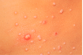
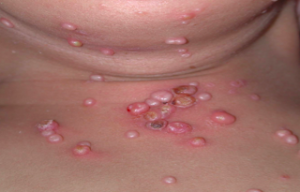
Molluscum Contagiosum (MC)

Screen Shot 2015-01-02 at 8.29.38 AM

Molluscum contagiosum usually occurs in children 0-14 years of age; however, Dr Friedlander reminds us that it can be seen in adults and transmitted sexually. The highest incidence occurs in children one to four years of age. There are definite associations that we need to keep in mind, i.e, swimming and eczema both appear to be associated with either a higher risk of getting the disease or a more prolonged course.

Unfortunately, there is a subset of patients may have a more prolonged or severe course.

Screen Shot 2015-01-02 at 8.29.59 AM

Families often want to know exactly how long it has been incubating and how long it will be there. Incubation is normally two to eight weeks. Molluscum infections can persist for years in some unlikely children, with an average duration of eight months. Generally, molluscum is asymptomatic; however, patients may be bothered with pruritus, erythema, bacterial superinfections, inflammation and pain.

What are the troublesome cutaneous findings associated with MC?

Atopy is prevalent in 44 percent of kids who present with MC (AD, asthma or allergies). What might be troublesome for you is the child who comes in with molluscum dermatitis, as this can occur in up to 39 percent of patients. The biggest problem is the patient with inflamed lesions as everyone thinks the child is infected and you need to take action. In one study, this occurred in 22 percent of patients. Another significant associated rash that we have seen is Gianotti-Crosti Syndrome-like reactions where you will see lichenoid papules around the elbows, knees, and buttocks. In children, if you note Gianotti-Crosti, type eruptions, think associated viral infections. Though we have traditionally been trained to think of EBV or hepatitis, Dr Friedlander suggests that you look to see if they have molluscum because many of them will.

What is BOTE?

BOTE is the “Beginning Of The End”, i.e., a predictor of resolution of the molluscum infection. Remember that the inflammatory phase of molluscum infection includes MC dermatitis, extreme induration and erythema, fluctuance, purulent exudate, “pseudo abcesses” and “ pseudo furuncles.” The onset of this type of inflammation to disease resolution is thought to be from three weeks to five months. (Butala N, et al. Pediatric. 2013;131:e1650.)

Why is it important to recognize “BOTE”?

The recognition of BOTE helps to avoid unnecessary cultures, antibiotics, admissions and the development of antibiotic resistance. Findings that should raise concern are a cellulitis-like appearance and lymphangitic streaking. If the child looks sick or is running fevers, you need to take action; however, if a child looks perfectly well, has multiple spots that have been red and inflamed looking for a while, and there’s no lymphangitic streaking, then you should keep BOTE in mind.

Treatment

What do the scientists tell us? Why is it so hard to clear? MC expresses proteins that circumvent immune responses (FLIPs) that inhibit b-interferon activation. What is Dr Friedlander’s hands down favorite therapy for molluscum? Cantheridin 0.7% in collodion applied with a Q-tip, washed off in four-to six hours. Some families are very nervous with this, so you can do test sites. Be sure to warn the family about the occurrence of blisters. What are the No-Nos? Catherone Plus for molluscum-NO. Do not do anything other than topical retinoids to the face , and intertriginous areas can develop severe blistering eroded reactions.

Cantharidin is an excellent treatment for molluscum but usually needs to be purchased from Canada or a compounded pharmacy. It can be ordered online through ABC pharmacy.com (Canthardin 7.5ml ~ $100.00) For those who have a willing hospital pharmacy, crystals can be ordered from Gallipot. The Cantharidin crystals themselves cost approximately $192.00/gm; when compounded it comes to $45.00/bottle.

Why don’t we let patients take Cantharidin home?

Topical application of Cantharidin for an extended time, extended areas or under occlusion and oral ingestion may cause serious side effects such as lymphangitis, TSS, and fatal poisoning. Beware of applying Cantharidin to intertriginous sites.

Another Option—The George Martin Method

  1. Obtain monochloracetic acid
  2. Fill small clean biopsy container half-way with crystals
  3. Add water half-way (saturated solution)
  4. Apply a very small amount with a toothpick
  5. Neutralize in 60 seconds with a wet paper towel

Patients can expect crusting and blistering in about two weeks.

Clinical Pearl—Remember the BOTE sign—avoid needless interventions in your molluscum patients

What do you need to think about when you see a purpuric rash?

You need to consider infection, i.e., bacterial, or viral. Also think about decreased production of marrow cells due to drug reaction, leukemia, or myeloproliferative disorders. Is there an increased consumption of marrow cells due to drug, ITP, hemolytic disorders or parvovirus? Let all of these possibilities run through your mind.

If you see lesions on the foot and hands, you may consider “hand, foot, and mouth disease” and Coxsackie. Usually Coxsackie virus is caused by A16. The disease usually occurs in spring-autumn in temperate climates and affects kids less than one to five years old. Most infections are asymptomatic. Patients may present with ulcerative stomatitis, lesions on the palms and soles and sometimes on the knees, elbows, buttocks, and genitals.

Coxsackie A6

Coxsackie 6 was first noted in 2011 by the CDC. How does it differ from regular Coxsackie? There is a wider age range with this disease. There may be vesiculobullous lesions present that are giant and a large area of involvement. There may be a rash that looks severe, but children tend not to look as sick. The problem with this disease is that if you were to try to culture it, it does not grow well in cultures. PCR however can nail it if it is available. You have to have clinical suspicion. What may help you nail the diagnosis if you don’t have PCR? Look at the mother’s nails.  And if a child comes in with nail shedding, think Coxsackie. Dr Friedlander’s practice saw several cases of onychomadesis in the office; when the moms were asked they mentioned that the children were sick about a month prior. Your first presentation of Coxsackie in the office may be a child whose nails are shedding or a mother’s.

What do we do with this child? We look at him/her, we look at the overall health of the child and we make sure that it isn’t something serious by clinical evaluation. When in doubt, evaluate for other more serious possibilities. With Coxsackie disease, we can reassure.

Look What the Wind Blew In…

A patient presents with a prolonged fever and a polymorphous eruption that looks a bit like erythema multiforme, a little bit like morbilliform, even measles-like. You need to think about bacterial and viral infections and you need to think about drug. You look at his conjunctivae and you notice limbal sparing, an area of white that is different from what you would get with adenovirus or Steven’s Johnson syndrome. When you undress the child, you notice a very distinctive perineal desquamating rash. This is Kawasaki’s Disease. You need to recognize this because while you may not see this often, when you do, you can save a life.

Kawasaki Disease (KD)

KD is a small- and medium-size artery vasculitis. There is now evidence that the incidence of KD may be linked to the velocity and direction of wind currents.

The typical age at presentation is six months to five years of age. The highest incidence of the disease is in Asian countries, and in the United States the incidence of KD is highest among the Asian population. If a child has KD, his/her siblings are six-to 30 times more at risk than the normal population and if a person had KD as a child, his/her child as a two time greater risk of the disease. The recurrence rate for the disease is two to four percent.

KD characteristics include a fever for more than five days as well as a rash that is variable but does not blister , and conjunctivitis (non exudative, non-limbal). KD affects the lips and tongue and there may be cervical lymphadenopathy as well as erythema/edema of the palms and soles as well as desquamation.

It is imperative that you intervene quickly with KD as this can decrease coronary risk by at least a factor of five.

Summary

With regards to pediatric infectious diseases, it is imperative that you know when to reassure, when to prevent unnecessary interventions and know when to act quickly!

Applying Laboratory Breakthroughs to Treat Pediatric Skin Diseases: Part 1

Amy Paller, MD, MS

At Maui Derm 2014, Dr Paller discussed how laboratory breakthroughs have enabled us to better treat skin disorders in the pediatric population. These discoveries come from the understanding of disease pathogenesis and our seeking of targeted interventions.

New discoveries and new technologies, e.g., gene sequencing, and analysis of RNA and protein expression, are increasing our understanding of skin disease and aiding in the development of new therapies.

Unraveling the Cause of Monogenetic Disorders

Discoveries from Sanger sequencing revolutionized prenatal diagnoses such as amniocentesis, chorionic villus sampling, and more recently for a limited set of disorders, maternal blood sampling. Preimplantation diagnosis is a technique that utilizes in vitro fertilization to pull out a single egg, extract the DNA, and distinguish whether or not the fertilized egg is normal or not and, thus, implant the eggs that will be normal.

Most dermatologists are familiar with Deep-Sequencing or “next-generation” sequencing technology, which is transforming our understanding of genetic diseases and how we can treat them.

Can next-generation sequencing help patients?

By simultaneously analyzing all candidate genes, we can detect low frequency mutations in known genes, mutations in newly associated genes, and low frequency SNPs in thousands of samples. We can also find genotype-phenotype correlations based on alterations in multiple genes. We can analyze epigenetic changes like methylation at base-pair level, histone modifications and protein-DNA interactions. This may provide us with a greater ability to predict responses to medications and personalized gene therapy.

Technologies in Trials Today

Gene therapy: Ex-vivo cell therapy

Researchers at Stanford are using this technology for recessive dystrophic epidermolysis bullosa (RDEB) in adults. One potential risk of ex vivo gene therapy is that the introduced protein may be viewed by the immune system as foreign, leading to a problem with the creation of autoantibodies. At Stanford, the researchers are only accepting patients (~60% of individuals with RDEB) who have the N-terminal domain of collagen VII—the immunogenic domain. This decreases the chance of the patient developing an autoimmune disorder. In these studies the concern about insertional oncogenesis (ie, that the viral insertion, which is random, could lead to interruption of an important tumor suppressor gene) is mitigated by the fact that any tumor that might developed would be highly visible and easily managed. On question is whether spraying the corrected keratinocytes onto a wound bed could be a substitute for grafting skin; this techniques has been used to introduce normal keratinocytes into graft sites for burn patients (Gerlach et al. Burns 2011;37:e19).

Stem Cell Therapy

Several years ago, the University of Minnesota began pioneering bone marrow transplants on children with RDEB, leading to evidence of incorporation of autologous stem cells, deposition of collagen VII at the dermal-epidermal junction, and some improved healing. Subsequent studies have used newer techniques to decrease the risk to patients and increase efficiency, including extending the therapy to junctional EB (JEB) patients. It is important to note that this procedure still carries a risk with variable success and slow improvement.

Revertant Mosaicism: “Natural” Gene Therapy

This term refers to a spontaneous correction of a loss-of-function mutation, leading to “carrier” phenotype. This has been seen extensively in patients with JEB because of mutations in collagen XVII, but has now been described in several other genetic disorders of the skin. This “natural gene therapy” may lead to expanding a small biopsy of patient cells to introduce as a graft or through use of iPS cells (see below) without requiring immunosuppression because the cells are otherwise the patient’s own cells.

Induced Pluripotent Stem Cells (iPS)

iPS cell technology allows one to take any cell (e.g., keratinocytes, fibroblasts) and de-differentiate it into a stem cell using well-known factors. You can then use other factors that will differentiate it into any type of cell that you want. As noted above, with regards to revertant mosaic areas, you can take those cells directly and turn them into stem cells and give someone back his own cells.

Many genetic diseases are dominant negative disorders, i.e., a genetic change in one allele is enough to disrupt function. Small interfering RNA (siRNA) is an approach to suppress the express of a target gene and has already been described in a human trial as a viable means to suppress gene expression and lead to clearance in pachyonychia congenita (Leachman et al. Mol Ther. 2010;18:442). siRNA specifically directed against the KRT6a mutation in a patient was injected into a tiny area of the plantar keratoderma using a left-right, double-blind analysis. Despite clear improvement, the injections were very painful, emphasizing the need for improved techniques for the delivery of genetic material through the epidermal barrier. Protrusion array devices utilize microneedles, often dissolvable, as a painless way to deliver genetic material or protein through the barrier. New creams that can be applied to the skin, including with the use of nanotechnology, are also under development as a means to deliver siRNA. TALENs and CRISPrs induce site-specific, double-stranded DNA breaks, then allow homologous-directed repair with the normal sequence. Although early in development, these techniques hold the promise of delivery of the normal sequence without the risk of random insertional oncogenesis.

Proteomic arrays (amino acid sequences encoded by mass spectrometry) and phosphoarrays (screens for activated proteins.) allow us to look at proteins directly and are great for drug discovery. ELISA assays have also become more sensitive. These technologies are allowing us to understand the specific pathways that are impacted in disease, leading to the possibility of targeted erapies for our patients with skin disease. With a better understanding of genes, we can develop newer therapies based on the pathways that they affect.


Melanoma Clinic: Lunch, Lesions, and Lessons: Part 4

Hensin Tsao, MD, PhD

Ilona Frieden, MD

Philip LeBoit, MD

Keith Flaherty, MD

Case Study

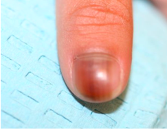
An 11 year-old boy presents to your office with his parents for the evaluation of a pigmented band on the right thumb. What would you do next?

Screen Shot 2014-12-05 at 2.46.54 PM

  1. Photo and follow up in 6 months
  2. Reassure and continue careful surveillance at home
  3. Perform an matrix biopsy with local anesthesia
  4. Matrix biopsy under general anesthesia

Melanonychia Management

Melanonychia is not rare; however, nail melanoma in children is rare. The pre-test probability of a benign outcome is high. Most of the time we can photograph and follow the patient over time. In an unscientific perusal of cases at UCSF, Dr Frieden found that the majority of the patients were of Asian descent. Occasionally, a biopsy is performed. In a 20-year cohort, there were no melanoma deaths in childhood from nail melanoma.

Special Sites and Concepts of “Mino”

Special sites, such as the nail, scalp, oral and genitalia, require careful scrutiny and some pathologists appear to have a better handle regarding the appearance of melanocytic lesions. The usual rules, both clinically and pathologically, often times do not apply. Definite cases of nail melanoma in childhood have been reported and there have been issues in the pathology of nail melanocytic biopsies. Richert and colleagues conducted a retrospective cohort of 30 biopsied lesions in Belgium. Seven out of the 30 patients were age 20 or younger and none of them had melanoma. (Richert B, et al. J Am Acad Dermatol. 2013;69(1):96-140.) A 2008 review article reported two cases of nail melanoma in children with skin type 3 and 4 in a 14 year-old and a 6 year-old. Both were diagnosed with having melanoma in situ (MIS) with onset of melanonychia less than one year of age. There were eight other reported cases in the literature with six diagnosed as MIS. Two had lymph node disease and neither presented as melanonychia. (Iorrizzo et al. Dermatol Surg. 2008;34:974-8)

“Struck by lightening versus severe bike accident?” Dr Frieden thinks about nail melanoma in terms of risk assessment. In general, we would not worry about being struck by lightening. It doesn’t mean it doesn’t happen, but it is extraordinarily rare.


Pediatric Dermatology: When to Suspect a Genetic Disease and How to Make a Diagnosis

James Treat, MD

In this presentation, Dr James Treat, an expert in pediatric dermatology, discusses genetic diseases in dermatology and provides us with information on how to make an accurate diagnosis.

Dr Treat reminds us that it is impossible to memorize all of the genetic diseases. As dermatologists, we can identify unusual cutaneous findings, i.e., describe the skin, as this can be helpful in making a genetic diagnosis. Remember that pathways can help you understand the disease(s). If you wonder about a genetic diagnosis, always ask yourself these four important questions:

  1. Does your patient have two or more unusual findings?
  2. Is there is a family history of similar findings?
  3. Is the child developing normally?
  4. Does the child have dysmorphic features?

Case Study Example

A child presents to your office with congenital red patches. What are they? What should we do about them?

  1. Does your patient have two or more unusual findings?—Yes, headaches
  2. Is there is a family history of similar findings?—Two uncles and his father have similar findings and he has an uncle with anyeurism
  3. Is the child developing normally?—He is having some difficulty in school
  4. Does the child have dysmorphic features?—No

If you have a patient who has multiple unusual findings OR multiple family members with one unusual finding, use your resources!

Technology provides us with an abundance of resources. When we have no idea or no specific search terms, we can use Google; however, we must be cautious in its use. In order to weed out non-scientific website answers, use Google Scholar. Does hand, foot, and mouth affect the buttocks? We can use Google images to search; yet, be aware that many images are mislabeled. OMIM can help when your patient has at least two unusual diagnoses and Genetests.org can aid if you’re wondering if a genetic test is available.

Capillary Malformation-Arteriovenous Malformation Syndrome (CM-AVM)

Dr Treat feels that this is something that is probably under-recognized. It’s a RASA-1 mutation that is associated with multiple capillary malformations that are inherited autosomal dominantly. There is usually a family history of multiple people with these red patches.

Clinical Pearls:

  • They look like café-AU-laits, but they are red-purple
  • They may all be nascent AVMS, so be careful using a laser

Remember that some patients have AVMs internally; they can be anywhere in the central nervous system and early screening for AVMs can be lifesaving. Given the association with CNS/spinal AVMs, the potential recommended work-up would be to consider an MRI/MRA.

Case Study Example

A patient presents with congenital red patches. He has a very large head with hydrocephalus. There is no other family history and he is developing normally. Does the child have dysmorphic features? Yes, he has a very large head and his 2nd and 3rd toes are fused (syndactyly). If we use our resources (Google Scholar or Google), we will find the name Macrocephaly-Capillary Malformation (and in small writing macrocephaly-cutis marmorata telangiectatica congenital CMTC).. M-CMTC was the name of this disease for a very long time, and went under-recognized because the skin finding is NOT CMTC it is a more recently described entity called a retiulated port wine stain. As dermatologists, we need to be able to tell the difference between the vascular formations.

Associations:

  • Macrocephaly
  • CM which tends to fade over time and tend to be more evanescent
  • 2nd-3rd toe syndactyly
  • Developmental delays

How do we diagnosis M-CM?

This disease was recently linked to disruption of the PI3k-AKT signaling. Findings indicative of M-CM are macrocephaly plus capillary malformations and two of the following:

  • Asymmetry of overgrowth
  • Developmental delay
  • Midline facial capillary malformations
  • Neonatal hypotonia
  • Syndactyly/polydactyly
  • Frontal bossing
  • Joint hypermobility
  • Hyperelastic skin, and hydrocephalus

This patient was diagnosed with hemangioma with arrested growth. How do we make the clinical diagnosis to differentiate hemangioma from capillary malformations and other vascular malformations? Look for red papules, ulceration (commonly seen in hemangiomas in this area), and large telangiectatic blood vessels as well as a peripheral rim of vasocontriction.

Case Study Example

Your patient presents with exuberant warts on the hands. With widespread, severe warts, we ask ourselves if there could be genetic immunodeficiency. Does your patient have other unusual findings? Yes, he has severe atopic dermatitis and always seems to be sick. There is no family history of similar findings, no dysmorphic features, and the child is developing normally.

Combined Immunodeficiency Associated with DOCK8 Mutations

DOCK8 was recently described as a cause of immunodeficiency that leads to severe viral infections, including the infections that we see as dermatologists, as well as upper respiratory tract infections and severe atopic dermatitis.

Most patients with severe atopic dermatitis and a few warts do not have anything wrong with their immune system; however, if you see a patient with terrible warts and terrible atopic dermatitis, you should think about having that patient see an immunologist.

Case Study Example

You notice congenital tan patches on your patient. What are they? Does your patient have other medical problems? No, and it’s only one tan patch. Is there a family history of similar findings? No. Is the child developing normally? Yes. Does the child have dysmorphic features? Yes, a large head.

This patient has neurofibromatosis Type 1 (NF1-). How do we diagnose this? We need two or more of the following: (Note that several of these findings are dermatologic)

  1. Six or more café-AU-lait spots 1.5cm or larger in post-pubertal individuals, 0.5cm or larger in pre-puberatal individuals
  2. Two or more neurofibromas of any type or one or more plexiform neurofibroma
  3. Freckling in the axilla or groin
  4. Optic glioma (tumor optic pathway)
  5. Two or more Lisch nodules (benign iris hamartomas)
  6. A distinctive bony lesion: dysplasia of the sphenoid bone or dysplasia or thinning of long bone cortex
  7. A first-degree relative with NF-1

What if you are sure that your patient has NF-1 and the testing is negative? You can also test for SPRED1.

SPRED1 is a novel mutation in the same pathway regulated by Neurofibromin, It is part of the SPRED/SPROUTY family which regulates MAP kinase activity (as does Neurofibromin). Most patients reported were adults and did not have other sequalae of NF-1. These patients have some increased risk of cognitive and developmental behavior abnormalities.

What is your workup in a healthy ten year-old with an isolated congenital flat tan patch?

  1. Send blood to evaluate for GNAS1 mutation
  2. Reassure
  3. Send blood to evaluate for NF-1 mutation
  4. Send blood to evaluate for SPRED1 mutation

These consults can be frustrating because we don’t always know what else can be doing on. Segmental pigmentary disorders (block-like, midline cutoff, isolated) have clinical characteristics that are reassuring as there is normally nothing else going on. Other diagnoses to be considered:

  • McCune Albright (typically multiple darker patches along thick lines of blaschko)
  • Neurofibromatosis (typically >6 oval smaller patches)
  • SPRED1 (same clinical café au lait macules as NF1)
  • Speckled Lentiginous nevi (by early childhood can often see the speckling within the larger tan patch)

Conclusions

You, as the dermatologist, have the power of accurate skin description. Look for capillary malformations, warts that are a bit too exuberant and café-AU-lait macules versus other pigmentary disorders. If you suggest a genetic disease, ask the important questions and utilize the resources that technology has provided us.


MauiDerm News Editor-Judy Seraphine

Pediatric Dermatology: Clinical Pearls

James Treat, MD

1.  Coxackie Virus can superinfect atopic dermatitis and look similar to eczema herpeticum

2.  Kwashiorkor can mimic severe atopic dermatitis in patients with severe nutritional restrictions

3.  Scabies almost never affects the face in children over two years of age

4. Mycoplasma can cause mucosal predominant stevens johnson with little to no skin lesions

5. Drug reaction with associated fevers are much more concerning and should be evaluated promptly

Eruption! Pediatric Acne

Larry Eichenfield, MD

Dr Eichenfield provided the audience at MauiDerm 2014 with an update on the new pediatric guidelines for the management of acne. These guidelines, published in May of 2013, were developed by the American Acne and Roseacea Society and endorsed by the American Academy of Pediatrics, which is the first time that the Academy has a set of guidelines for the management of acne. One of the main reasons this was done was to address the practice gap between dermatologists and pediatricians. The group of experts was comprised of dermatologists with expertise in acne, pediatric dermatologists, and pediatricians.

As we know, acne ranges in terms of presentation and severity.  Acne can be categorized by age, i.e., neonatal (0-6wk), infantile (0-1), mid-childhood (1-7), preadolescent (7-12), and adolescent (12-19).

Dr Eichenfield emphasizes that mid-childhood acne (age 2 – 7) acne is the most worrisome. It can be associated with premature adrenarche, Cushings Syndrome, CAH, gonadal/adrenal tumors, and precocious puberty. Mid-childhood acne is very uncommon, and is a sign of early adrenarche, often associated with a pathologic process. The assessment for mid-childhood acne includes assessment of  testicular size (males), presence or absence of hirsutism, clitoromegaly, androgenetic alopecia, increased muscle mass, and/or deepening of the voice (males).  Tests and examinations include a growth chart, evaluation including bone age, tanner stage, Total/Free testosterone, DHEAS, androstenedione, LH, FSH, prolactin, and 17OH-progesterone. The guidelines suggest that you should refer these patients to a pediatric endocrinologist.

What do the new guidelines say?

It’s important to know that pre-adolescent (7 ≤ 12 years) acne is common and may precede other signs of pubertal maturation. Work-up beyond history and physical is generally unnecessary unless there are signs of androgen excess, polycystic ovarian syndrome, or other systemic abnormalities. According to the data, there is evidence that acne, and possibly puberty, are now occurring at an increasingly earlier age for both males and females. If you look at some of the papers that were published about a decade ago, the amount of comedones that were seen in a ten or 12 year-old then is probably different now. Twelve is no longer an age point defining “normal acne;”  if you are eight and above acne can be typical and common.

After the guidelines were published early acne in preteens was highlighted in several newspaper articles, and radio and television segments.  Articles were published in both the New York Times and USA Today. In fact, after the New York Times article was published, there were over 157 blogs that provided an interesting perspective comparing what the general public thinks regarding acne and what we do as specialists.  A recent analysis of the National Ambulatory Medical Care Survey (NAMCS) database assessed trends in the age of children seeking treatment for acne. NAMCS data from 1979 through 2007 were analyzed for all physician visits in which acne vulgaris was listed as a diagnosis in children aged 6 to 18. The analysis revealed a significant decrease in the mean age of children seeking treatment for acne over this period.

Over the past several years, clinicians have noted an earlier onset of acne—that is, the appearance of acne in patients as young as 8 or 9 years. Indeed, it has been suggested that 12 years of age should no longer be considered the low end of the “normal” range for the onset of acne.

Acne may be the first sign of the onset of puberty in children aged 7 to 11 years. The general clinical impression of earlier puberty is, in fact, supported by epidemiologic data in the US and elsewhere. In general, the trend toward earlier puberty is stronger among girls than among boys. In any event, the earlier onset of acne has mirrored the downward trend in puberty timing.

The guidelines provide an algorithm for the management of pediatric acne. If you can generally categorize the acne as mild, moderate or severe, you can access the algorithm. Keep in mind that this algorithm is slightly different than prior guidelines, in that for mild acne initial treatment benzoyl peroxide, based upon the evidence, made it as one potential, initial solo therapy.   The guidelines can be found in the Journal of the American Academy of Pediatrics, or online at: http://pediatrics.aappublications.org/content/131/Supplement_3/S163.full.pdf.

Acne Guidelines: Highlights

The guidelines emphasize appropriate use of medications based upon disease severity.  Oral antibiotics should be used concurrently with a topical retinoid because it is important to build a topical regiment to “transfer the patient to” after a limited course of antibiotics.   A variety of studies show that 70 percent of the time you can transition your patient who is on an oral antibiotic and topical retinoid to a regiment of topical retinoid alone or combinations with topical antimicrobials (like benzoyl peroxide) and/or antibiotics

Topical retinoids may be used as monotherapy or in combination products and in regimens of care for all types and severities of acne in children and adolescents of all ages. It is important to remember that topical antibiotics are not recommended as monotherapy and if a topical antibiotic treatment is to be used for more than a few weeks, topical benzoyl peroxide should be added, or used in combination products. The guidelines also suggest that fixed-dose combination topical therapies may be useful in regimens of care for all types and severities of acne.

With regards to oral antibiotics, they are a reasonable approach for moderate to severe inflammatory acne vulgaris at any age. Tetracycline derivatives should not bee utilized in children 8 years of age and below. Second generation tetracyclines are “sometimes preferred” to tetracycline because of ease of use, fewer problems with absorption with food and minerals in vitamins and other supplements, and less frequent dosing. It’s very important that patients are educated and monitored for potential adverse events when utilizing oral antibiotics for acne.

Hormonal therapy is actually interesting because there are people who are very pro-hormonal therapy and others are a bit more conservative and prefer oral antibiotics. The group ended up stating that combined oral contraceptives (OCs) may also be useful as second-line therapy in regimens of care in pubertal females with moderate to severe acne; however, tobacco use and family history of thrombotic events should be assessed. Due to concerns about growth and bone density, many recommend withholding OCs for acne until one year after onset of menstruation.

Isotretinoin is recommended for severe, scarring, and/or refractory acne in adolescents and may be utilized in younger patients. Remember that extensive counseling, particularly regarding the avoidance of pregnancy, as well as careful monitoring of potential side effects and toxicities, is recommended. As far as the specific discussion on isotretinoin in pediatric use, there are bone mineralization changes; however, the data is inconsistent and it is not associated with increased factures. Hyperostoses are very uncommon for acne and there has been one case of premature epiphyseal closure in a patient on isotretinoin for acne, but that’s not necessarily attributable to the isotretinoin. IBD is controversial, but counseling is reasonable.

Practice Gaps

Dr Eichenfield states that there is a chasm in the way that General Practitioners, Pediatricians, and Dermatologists treat acne. A 2014 paper by Tan et al showed that topical retinoids were prescribed in 41 percent of acne visits. Older age, male gender and having Medicaid insurance were associated with a lower likelihood of receiving a topical retinoid prescription. Moreover, the researchers found that in the Medicaid dataset, patients who saw a pediatrician or general practitioner had lower odds of receiving a topical retinoid prescription versus those patients who saw a dermatologist.

Another study looked at the National Ambulatory Medical Care Survey (NAMCS) data regarding the treatment of preadolescent acne in the United States. The data were stratified according to age group and physician specialty. The findings are presented below:
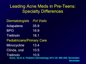
Screen Shot 2014-06-11 at 1.39.02 PM


What are Dr Eichenfield’s thoughts on the practice gaps?

  • Over-reliance on oral antibiotics
  • Use of oral antibiotics without BP
  • Use of oral antibiotics without Retinoid
  • Use of topical and oral antibiotics together, without retinoid
  • Under appreciation of early, significant acne as predictor of worse acne over time

Literature has shown that early comedonal acne may predict later severe acne. So remember that if you see a patient with a high number of comedone counts at an early age, their chances of severe inflammatory acne at a later age is much higher than someone who has low comedone counts early on.

Another issue that Dr Eichenfield is appreciating more and more in clinical practice is that you can have very early, subtle scarring. We know that there is a lot of scarring that occurs without nodular-cystic disease, so this is very common. This is an important to try to get patients evaluated early so that scarring can be prevented, and minimized. Dr Eichenfield advised  that a useful technique is to side-light the face and look for depressions in the face, displaying  scarring as opposed to post-inflammatory hyperpigmentation or persistent erythema.

A study by Patel and colleagues aimed to determine what types of acne lesions preceded the development of atrophic acne scars. Twenty-two patients with mild to moderate acne were enrolled in a split-face study in which one side was treated with non-ablative laser and the other remained untreated. A series of standardized digital facial photographs was obtained from the untreated side at 2-week intervals from baseline to week 12, and all photographs in the series were aligned with the baseline photo. When all of the atrophic scars were tracked to baseline, 53 were found to have arisen from clinically normal skin, 30 were established scars, and 21 arose from acne lesions, including closed comedones. However, no open comedones at baseline corresponded to atrophic scars.  The results of this study not only verify that inflammatory acne lesions often lead to atrophic scarring, but also demonstrate that acne scars may arise from initially comedonal lesions, as well as from clinically normal skin. Moreover, they indicate that a period of 12 weeks is sufficiently long to develop and establish atrophic scars. Thus, aggressive treatment of both inflammatory and comedonal acne is warranted to minimize acne scarring.

What about Isotretinoin?

We know that isotretinoin is the most effective treatment for acne; however, the optimal dosing regimen is still unknown.  Dr Eichenfield comments that he uses isotretinoin commonly. He also states that he tends to be biased towards lower-dose isotretinoin on a daily basis, working up to cumulative doses of 120-150 mg/kg.  He commonly will the daily doeses up to the “highest comfortable dose,”  that is, the highest dose with minimal significant side effects or laboratory abnormalities.

A recent publication looked at 180 patients with acne resistant to other treatments who were enrolled in an observational, prospective study of istotretinoin with cumulative doses less than to 220 mg/kg versus isotretinoin greater than or equal to 220 mg/kg. Of these patients, 116 participated in the 12-month follow-up survey. At that time, 97.4 percent of the patients reported that their acne was improved. Overall, acne in 32.7 percent of the patients in the study relapsed at 12 months, and 1.72 percent of the patients required a retrial. In the lower-dose treatment group, the relapse rate was 47.4 percent compared with 26.9 percent in the high-dose group. Almost 100 percent of the patients in both treatment groups developed cheilitis and xerosis during treatment. Retinoid dermatitis was significantly more common in the high-dose treatment group and none of the other adverse effects were significantly different between the two groups.  However,  it should be noted that in the higher dose group,  nine patients had  persistent muscle aches, eight patients had persistent joint aches, and two patients  had hearing changes. (Blasiak RC et al. JAMA Dermatol 2013;149(12):1392-1398) Also of importance with regards to this study are the laboratory abnormalities based upon the dosing. (See table 1)
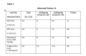
Screen Shot 2014-06-11 at 1.39.15 PM


Dr Eichenfield states that this publication has yet to “move him” to abandon his current methodology with regards to isotretinoin dosing for this patients.

Idiopathic Facial Asceptic Granuloma (IFAG) and Childhood Rosacea

We have seen a very interesting change in perspective regarding this disease. Occasionally, we see these patients who present with lumpy, cystic-type lesions, separate from acneiform lesions.   A multi-center study of four French dermatologic centers looked at patients who were diagnosed with IFAG between October 2000 and July 2007. Thirty-eight patients were included in the study. The median age at the time of diagnosis of IFAG was 43 months, with a median follow-up of 3.9 years. Sixteen patients (42.1%) had at least two criteria of childhood rosacea, 11 of 32 (34.4%) with a single lesion and 5 of 6 (83.3%) with multiple lesions. Remember that childhood rosacea presents with flushing, permanent or recurring erythema, papules and postules without comedones or microcysts, convexity predominance of lesions, ocular rosacea (chalazions, conjunctival hyperemia, keratitis). (Prey S, Ezzedine K et al. Pediatric Dermatology 2013;30:429-32)

What does this mean to us? Children with IFAG are at risk for childhood rosacea, and follow-up is advised, including periodic ophthalmologic assessment.


MauiDerm News Editor- Judy Seraphine