Melanoma Clinic: Lunch, Lesions and Lessions: Part 2

Part 2

Hensin Tsao, MD, PhD

Keith Flaherty, MD

Ilona Frieden

Philip LeBoit, MD

Case 2

In early August of 2007, a 47 year-old patient presented to her PCP with concerns of an atypical appearing lesion on the left lower leg. She was referred to a dermatologist. On August 14th, the biopsy showed 0.97 mm, Level IV, SSM, four mitoses, and non-ulcerated. A wide excision was performed and there was no residual melanoma and no further surgery was recommended. ‎In April of 2009, the patient noted a small cutaneous mass in scar line on the lower left leg.  In June, the patient underwent wide local excision and sentinel lymph node biopsy. The results showed two out of three positive sentinel nodes, the largest metastasis was 4mm; and (+) extracapsular extension.  On July 29, 2009, she underwent (L) inguinal LND and sartorius muscle transposition flap.  Zero out of seven nodes (including Cloquet’s node) were negative.   There were a total of two out of ten positive nodes. She received adjuvant interferon. Unfortunately, two years later, a PET-CT showed an increase in the number of FDG avid lesions in the leg and inguinal and pelvic adenopathy. She’s now considering systemic treatment.

What specimen should be used for determining the patient’s BRAF status?

  1. Primary melanoma FFPE
  2. Primary melanoma FFPE and blood DNA for comparison
  3. Sentinel lymph node metastasis
  4. Soft tissue metastasis from leg

The general guidelines are to take the most recent evidence of advanced disease. Concordance is very important. Do you know that the lesion in the lung is identical to the lesion that presented five years ago according to its genotype?

SNaPshot panel reveals BRAF WT, NRAS and Q61R mutation. On March 21, 2011, she received high-dose IL-2 therapy. In June of 2011, the PET/CT revealed increased FDG uptake in pre-existing left leg subcutaneous nodules and an increase in size as well as FDG uptake in bilateral inguinal lymph nodes. In August, she enrolled in a clinical trial of bevacizumab and ipilimumab. A CT C/A/P performed in April of 2012 revealed an increase in the size of the largest lung mestastasis (15mm to 20mm) and a new 1.6cm liver metastasis. MRI of the brain was negative. June 8, 2012 was her first day on 10-017, a phase 1 study of a CDK4/6 dual inhibitor. By the end of the July, CT C/A/P revealed disease progression in the lung and a new, solitary brain metastatsis measuring 4mm; the disease burden was otherwise relatively stable. In August, she underwent stereotactic radiation to the solitary, 4mm brain mestastasis. October 23, 2012 was cycle one/day one of BKM120/MEK162 trial # 11-308. She remained stable for four months.

Screen Shot 2014-10-26 at 8.18.34 PM

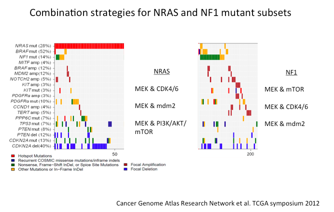
One of our greatest challenges is that NRAS is not something that we can target with a drug. Indirect strategies; however, may work.

Screen Shot 2014-10-26 at 8.18.48 PM

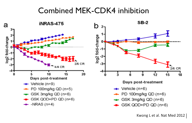
MEK inhibitors in NRAS-mutant melanoma have some efficacy. It’s not comparable to what a MEK inhibitor or BRAF inhibitor can do in BRAF-mutant cancer, but it is certainly a suggestion that may perturb these tumors.

Contact Dermatitis

Matthew Zirwas, MD

Dr Matt Zirwas provides us with his top ten take-home points from his workshop at MauiDerm NP+PA in Boston…


  1. Add 10 allergens to the TRUE Test to improve diagnostic reliability dramatically.
  2. OK to patch test on 10 mg of prednisone and all the antihistamine in the world.
  3. OK to patch test on immunosuppressives as long as they still have some rash.
  4. Final read MUST be between 96 and 120 hours after patches are put on.
  5. When reading patches, what you feel is what matters, not what you see.
  6. Most positive patch test reactions don’t matter and you shouldn’t even tell the patients about them.
  7. Negative patch testing is extremely helpful.
  8. If have relevant patch test results, must avoid information overload – tell them the minimum amount of information necessary.
  9. Telling people what they CAN use is much more important that telling them what they can’t use.
  10. Occupational cases almost never turn out well.

Vasculitis and Vascular Lesions Part 1: Clinical Pearls

Ted Rosen, MD

1. Vasculitis is a clinicopathologic process characterized by inflammation of and damage to blood vessels
2. Heterogeneous group of disorders leading to a broad group of named diseases due to….  Different sizes, types and locations of the affected blood vessels and variable tissue response to injury
3.  Basic principles:

  • All age groups (Some types have preference)
  • Caucasians (But other ethnic groups, too)
  • Genetic predisposition (But not strictly inherited)
  • Chronic, relapsing (But may go into remission)
  • Think vasculitis when faced with an…..unexplained multi system disease or progressive organ dysfunction
  • Team approach: Early consultations

4. Classification: best done by size of blood vessel affected

5. A wide variety of tissues can be affected. Scoring charts exist which are helpful to systematically asses these patients.

6. Constitutional signs and symptoms may be seen: fever, chills, sweats, weight loss, fatigue

7. Involvement of, damage to and symptoms relating to: eyes, upper or lower airway, joints, kidneys, lungs, central/peripheral nervous system,  heart and clotting mechanisms

8. Morphology of skin lesions is suggestive but virtually never diagnostic of histologic type!

9. Absence of skin lesions does not rule out any variant of vasculitis, while the presence of any specific type of skin lesion does not necessarily predict the subtype of vasculitis present!

10. There are many clinical mimics to vasculitis. These include embolic phenomenon, exposure to certain drugs, cryptogenic infections, Vitamin C deficiency, and cardiolipin antibody syndrome

11. When biopsy is done, try to utilize a lesion less than 72 hours old to get the most representative histology.

12. Therapy may include: NSAIDS, Dapsone, Colchicine, Steroids, Immunosuppressives: Methotrexate, Mycophenolate, Azathioprine, Cyclophosphamide and Rituximab

Infectious Disease Update: Clinical Pearls

Ted Rosen, MD

At MauiDerm Fall NP+PA 2014, Dr Rosen provides us with an update on infectious diseases and his clinical pearls…

  1. STDs are globally epidemic. In the US, 50% of those 15-25 have or have had an STD
  2. STDs are so common due to the “multiplier” effect of sex partners of sex partners
  3. Syphilis is on the rise last three years in USA, now at 5.3/100,000 population
  4. Multi-drug resistant GC (including resistance to drugs of choice, cephalosporins) is here
  5. Topical three months regimen of 4% zinc sulfate soaks for active genital herpes benficial
  6. External genital warts: NEW treatment ideas
    • 5% KOH daily x 3 months
    • Single application of ingenol mebutate (either strength)
    • Thermotherapy with 440C for 30 min x 3 days, each of two consecutive weeks
  7. Leishmaniasis being brought back to USA from Iraq and Afghanistan
    • Rx: Pentavalent antimonial IV, IM from CDC; Quite toxic.
    • Thermotherapy (ThermoMed device): 1300F; Painful and blisters
    • New drug: Miltefosine 100-150mg/day PO (Impavido™) available 2015
      • Lecithin derivative
      • Safe and effective for Old and New World Leishmaniasis
  8. Treatments for MRSA have expanded with three new drugs
    • All new drugs have LONG T1/2 which allows less frequent dosing
    • Oritivancin: Single 1200mg dose
    • Dalbavancin: 1000mg, then 500mg a week later
    • Tedizolid: 200mg QD x 5-7 days
  9. Chikungunya virus
    • Used to be Africa/Asia, Now worldwide
    • Most cases imported from trip (especially from Caribbean)
    • Now, virus-carrying mosquitoes in Caribbean and USA
    • Fever, arthralgia, myalgia, headache, petechial to purpuric rash
    • Like Dengue but not as bad
    • No vaccination, No specific Rx. Supportive therapy, as needed.

Melanoma Clinic: Lunch, Lesions, and Lessions

Part 1

Hensin Tsao, MD, PhD

Keith Flaherty, MD

Ilona Frieden

Philip LeBoit, MD

Dr Hensin Tsao, of Harvard Medical School, discusses several challenging cases with regards to the management of melanoma and pigmented lesions…Remember that melanoma, as an area, is not always easy.

Case 1

A ten year-old girl presents with a pink plaque on the left shoulder. The lesion has been present for four months and she has no other medical problems.

Screen Shot 2014-08-24 at 3.10.15 PM

As a clinician, what would you do next?

  1. Photo and follow up in 6 months
  2. Reassure and continue careful surveillance at home
  3. Perform an excisional biopsy with local anesthesia
  4. Biopsy under general anesthesia

Why not photograph and follow or reassure and continue surveillance. Because of the shape of this lesion, it’s a little bit bigger than normal spitz nevus. Because of her age, she can probably tolerate anesthesia so it would be worth obtaining a good specimen. The biopsy showed 1.5mm thick, Clark level IV, it was non-ulcerated and 2 mits/mm2.

Managing Pediatric Melanoma

A study by Cordoro and colleagues demonstrated that pediatric melanoma does not follow the ABCDs. What do we look for? Most teenagers who get melanomas get adult-type melanoma. Up until age 13, melanoma is diagnosed at a relatively low level. Amelanotic or bleeding is also more frequent presentations of melanoma in younger kids.

Screen Shot 2014-08-24 at 3.10.28 PM

ABCDE Modified for Children

  • Amelanotic
  • Bleeding; bumps
  • Color uniformity
  • De Novo; Diameter; any
  • Evolution

Make sure that you have a dermatopathologist with whom you are very confident!

Risk stratification, rather than black and white diagnosis, is the way things are going now with regards to tumors and general pathology. There are lesions where we cannot always make a precise diagnosis, even after molecular testing. In many of these cases, all we can say is that it is a Spitzoid neoplasm with very low or high malignant potential. Spitzoid neoplasms can be stratified into four categories. These are Spitz nevus, Spitz nevus with atypical features, atypical Spitz tumor and Spitzoid melanoma.  Bastian and his colleagues have developed Spitzoid taxons:

  • H-ras mutated
  • Bap-1 mutated
  • Braf fusion
  • Alk fusion
  • Ntrak1 and ntrak3 fusion
  • Ros-1 fusion

One very interesting concept is that many of these genes, if you take them and translocate them in cells other than melanocytes, they are extremely potent in terms of oncogenic effect. ROS-1 and Alk, for example, are important players in lung cancer. Mutations or fusion partners do not define benign versus malignant, but only lineage. So, doing a test for Alk will not tell us if the lesion is benign or malignant; however, if the lesion does metastasize these are very important venues for therapy. For instance, BRAF, even though there’s no Braf mutation in these lesions, it is over-expressed and anti-BRAF therapies may be effective in Spitzoid melanomas that have a BRAF fusion. You don’t have to have the mutation for BRAF inhibition to work, if it’s a tumor where other things are turning it on.

Screen Shot 2014-08-24 at 3.10.49 PM


Systemic Medications: Top Ten Take Home Points

Matthew J. Zirwas, MD 

  1. Prednisone is drug of choice for expected short term therapy – weeks.
  2. Cyclosporine is drug of choice for expected “medium term therapy” – months.
  3. Methotrexate is drug of choice for chronic therapy in patients without liver, kidney, or hematologic co-morbidities.
  4. Mycophenolate is drug of choice for chronic therapy in patients with liver, kidney, or hematologic co-morbidities.
  5. Cyclosporine has many drug interactions – must use a computerized drug interaction checker.
  6. Methotrexate has a few drug interactions but they are extremely serious and can be fatal.
  7. Mycophenolate has few drug interactions.
  8. Patients on methotrexate should take folic acid on days they do not take the methotrexate.
  9. Older patients on mycophenolate, in particular, are at high risk of zoster.
  10.  If patients on cyclosporine have elevations in creatinine, they need to stop the medication.

Pigmented Lesions: Clinical Pearls

Ashfaq Marghoob, MD

Dr Marghoob provides us with his clinical pearls on pigmented lesions….

  1. The larger the congenital nevus the greater is the risk for developing melanoma.
  2. The risk of a melanoma developing in a small congenital nevi is small enough that prophylactic excision is not required.
  3. Presence of many nevi and dysplastic nevi are strong risk factors for melanoma.
  4. Analytical, Differential and comparative recognition are all helpful in differentiating nevi from melanoma.
  5. Total body photography assists clinicians in finding concerning lesions.
  6. Dermoscopy assists clinicians in deciding which lesions require a biopsy.
  7. Unna’s theory of nevogenesis is not supported by recent cross sectional and longitudinal studies.
  8. Nevi with a peripheral globular pattern or a starburst pattern are growing nevi (not yet in senescence).
  9. Most halo nevi have a globular pattern.
  10. Patient driven healthcare with the use of Apps is likely to help in finding early melanomas.

Immunology 101: The Basics

Andrew Blauvelt, MD

Dr Blauvelt provides us with the 10 most important take-home messages from his immunology presentation at MauiDerm NP+PA Summer 2014….

  1. Key features of the innate immune system include: rapid response, non-specificity, phagocytosis, no memory.
  2. Key features of the  acquired immune system include: slow response, very specific, lymphocyte-mediated, memory.
  3. Keratinocytes are active participants in generating immune responses by secreting numerous cytokines upon activation.
  4. 4.Toll-like receptors are pattern recognition receptors on keratinocytes that recognize foreign antigenic material.
  5. Antimicrobial peptides are natural antibiotic molecules found in skin that are abundant in psoriasis skin and sparse in atopic dermatitis skin.
  6. 6.Epidermal Langerhans cells are antigen presenting cells that recognize/process skin antigens and migrate to lymph nodes, where they present antigen to T cells.
  7. 7.T cells require 3 signals to become fully activated: 1) recognition of antigen by the T cell receptor via MHC on the surface of antigen presenting cells; 2) binding of co-stimulatory molecules on T cells and antigen presenting cells to one another; and 3) secretion of soluble cytokines by the T cells.
  8. CD4+ T cells are T helper cells that recognize antigen via MHC class II and secrete cytokines to enhance CD8+ T cell and B cell immune responses, whereas CD8+ T cells are cytotoxic T cells that recognize antigen via MHC class I and kill cells upon contact.
  9. B cells secrete antibodies that specifically bind to antigen.
  10. Primary immune responses are slow, occur after first exposure to antigen, and involve creation of memory cells, whereas secondary immune responsesare fast, occur after subsequent exposures to antigens, and involve reactivation of memory cells.


Dermatitis Overview: 2014

Matthew J. Zirwas, MD

Are you treating dermatitis? Dr Zirwas provides us with ten clinical pearls…

  1. The main goal in dermatitis patients is figuring out WHY they have dermatitis.
  2. Acute allergic contact dermatitis starts 1-3 days after exposure to the allergen and takes 1-3 weeks to go away.
  3. Chronic allergic contact dermatitis takes 3-6 months to go away.
  4. Hypoallergenic rubber gloves are not hypoallergenic.  Hypoallergenic gloves include: vinyl gloves, Microtouch Nitra-Free, Dermaprene Ultra.
  5. Shampoo and conditioner are the main causes of eyelid dermatitis, both irritant and allergic.
  6. Atopic dermatitis is the combination of inadequate skin barrier and an immune system that was genetically primed to go in an allergic direction.
  7. Food avoidance and chelation do not work in adults.
  8. There are probiotics that work, but it is very specific – patients can’t just take any old probiotic.
  9. Seborrheic dermatitis is an inflammatory reaction to Malassezia yeast on the skin – treat by reducing amount of yeast and targeting the inflammation.
  10. Xerosis is too little water in the skin.  Need to add water and prevent it from just evaporating away.


Dermoscopy: Clinical Pearls

Ashfaq Marghoob, MD

Are you performing dermsocopy in your clinical practice? Dr Marghoob provides us with his clinical pearls…

  1. Dermoscopy helps in differentiating benign lesions from skin cancer.
  2. The presence of network (reticular network, negative network, polygonal lines), streaks (radial streaming, pseudopods), aggregated globules/peripheral globules, homogeneous blue pigmentation is most often seen in melanocytic lesions.
  3. Melanocytic lesions that deviate from the 10 benign patterns and has at least one of the 10 melanoma-specific structures needs to be biopsied to rule out melanoma.
  4. The presence of spoke wheel structures and/or leaf like areas is 100% specific for BCC.
  5. Comedo like openings and milia cyst are often seen in SK but can also be seen in other lesions including melanoma.
  6. Polarized light makes blood vessels and crystalline structures more conspicious, and makes milia cyst less conspicuous.
  7.  A polymorphous vascular pattern in an amelanotic lesion should raise concern for melanoma.
  8. Dermoscopy improves the clinician’s diagnostic accuracy.
  9. All structureless or featureless or not-otherwise-diagnosable lesions should be viewed with suspicion.
  10. Raised lesions should never be monitored for change.